• 文献检索
  • 文档翻译
  • 深度研究
  • 学术资讯
  • Suppr Zotero 插件Zotero 插件
  • 邀请有礼
  • 套餐&价格
  • 历史记录
应用&插件
Suppr Zotero 插件Zotero 插件浏览器插件Mac 客户端Windows 客户端微信小程序
定价
高级版会员购买积分包购买API积分包
服务
文献检索文档翻译深度研究API 文档MCP 服务
关于我们
关于 Suppr公司介绍联系我们用户协议隐私条款
关注我们

Suppr 超能文献

核心技术专利:CN118964589B侵权必究
粤ICP备2023148730 号-1Suppr @ 2026

文献检索

告别复杂PubMed语法,用中文像聊天一样搜索,搜遍4000万医学文献。AI智能推荐,让科研检索更轻松。

立即免费搜索

文件翻译

保留排版,准确专业,支持PDF/Word/PPT等文件格式,支持 12+语言互译。

免费翻译文档

深度研究

AI帮你快速写综述,25分钟生成高质量综述,智能提取关键信息,辅助科研写作。

立即免费体验

超分辨率荧光显微镜成像的核纤层蛋白的定量分析。

Quantitative Analysis of Nuclear Lamins Imaged by Super-Resolution Light Microscopy.

机构信息

Department of Cell and Molecular Biology, Feinberg School of Medicine, Northwestern University, Chicago, IL 60611, USA.

Department of Biophysics, UT Southwestern Medical Center, Dallas, TX 75390, USA.

出版信息

Cells. 2019 Apr 18;8(4):361. doi: 10.3390/cells8040361.

DOI:10.3390/cells8040361
PMID:31003483
原文链接:https://pmc.ncbi.nlm.nih.gov/articles/PMC6524165/
Abstract

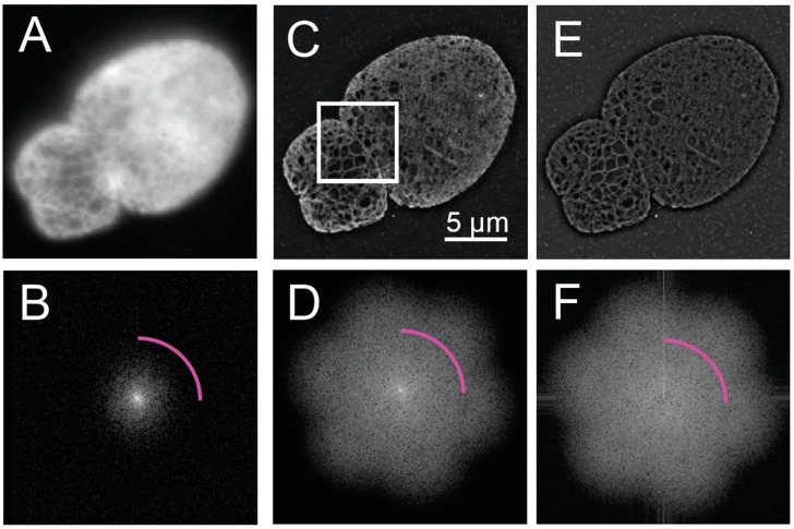
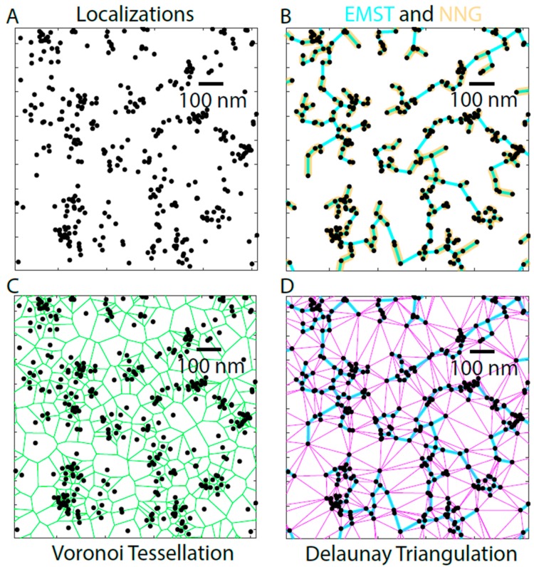
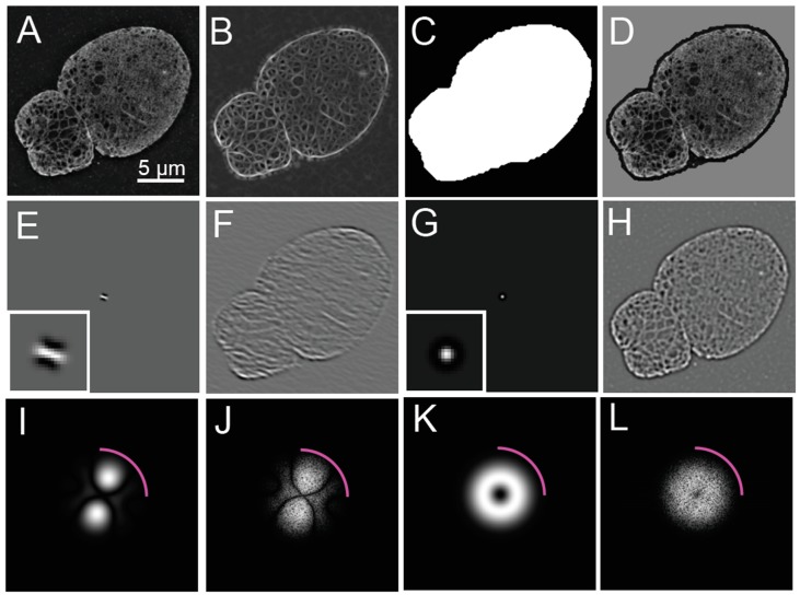
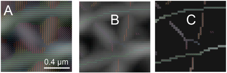
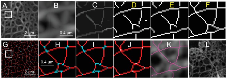
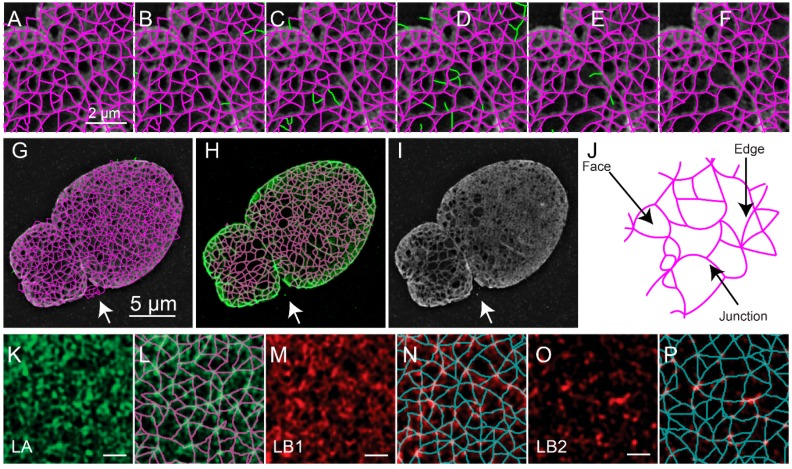
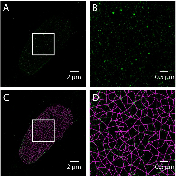
The nuclear lamina consists of a dense fibrous meshwork of nuclear lamins, Type V intermediate filaments, and is ~14 nm thick according to recent cryo-electron tomography studies. Recent advances in light microscopy have extended the resolution to a scale allowing for the fine structure of the lamina to be imaged in the context of the whole nucleus. We review quantitative approaches to analyze the imaging data of the nuclear lamina as acquired by structured illumination microscopy (SIM) and single molecule localization microscopy (SMLM), as well as the requisite cell preparation techniques. In particular, we discuss the application of steerable filters and graph-based methods to segment the structure of the four mammalian lamin isoforms (A, C, B1, and B2) and extract quantitative information.

摘要

核纤层由核纤层蛋白、V 型中间丝等组成,呈密集的纤维网格状,根据最近的冷冻电镜断层扫描研究,其厚度约为 14nm。近年来,光学显微镜技术的进步将分辨率扩展到了一个可以在整个细胞核背景下对核纤层精细结构进行成像的尺度。我们综述了通过结构光照明显微镜(SIM)和单分子定位显微镜(SMLM)获取核纤层成像数据的定量分析方法,以及必要的细胞制备技术。特别是,我们讨论了可转向滤波器和基于图的方法在分割四种哺乳动物核纤层蛋白(A、C、B1 和 B2)结构和提取定量信息方面的应用。

https://cdn.ncbi.nlm.nih.gov/pmc/blobs/b942/6524165/69b947823898/cells-08-00361-g007.jpg
https://cdn.ncbi.nlm.nih.gov/pmc/blobs/b942/6524165/c50268bb64db/cells-08-00361-g001.jpg
https://cdn.ncbi.nlm.nih.gov/pmc/blobs/b942/6524165/0da2de731409/cells-08-00361-g002.jpg
https://cdn.ncbi.nlm.nih.gov/pmc/blobs/b942/6524165/c6eadcb7538a/cells-08-00361-g003.jpg
https://cdn.ncbi.nlm.nih.gov/pmc/blobs/b942/6524165/0ca92e054894/cells-08-00361-g004.jpg
https://cdn.ncbi.nlm.nih.gov/pmc/blobs/b942/6524165/471faccb9735/cells-08-00361-g005.jpg
https://cdn.ncbi.nlm.nih.gov/pmc/blobs/b942/6524165/f394545b70d4/cells-08-00361-g006.jpg
https://cdn.ncbi.nlm.nih.gov/pmc/blobs/b942/6524165/69b947823898/cells-08-00361-g007.jpg

相似文献

1
Quantitative Analysis of Nuclear Lamins Imaged by Super-Resolution Light Microscopy.超分辨率荧光显微镜成像的核纤层蛋白的定量分析。
Cells. 2019 Apr 18;8(4):361. doi: 10.3390/cells8040361.
2
The molecular architecture of lamins in somatic cells.体细胞中层粘连蛋白的分子结构。
Nature. 2017 Mar 9;543(7644):261-264. doi: 10.1038/nature21382. Epub 2017 Mar 1.
3
Nonlinear mechanics of lamin filaments and the meshwork topology build an emergent nuclear lamina.层状细丝的非线性力学和网格拓扑结构构建出一个新兴的核层。
Nat Commun. 2020 Dec 4;11(1):6205. doi: 10.1038/s41467-020-20049-8.
4
Structural and physiological phenotypes of disease-linked lamin mutations in C. elegans.线虫中与疾病相关的核纤层蛋白突变的结构和生理表型。
J Struct Biol. 2012 Jan;177(1):106-12. doi: 10.1016/j.jsb.2011.10.009. Epub 2011 Nov 7.
5
Structural organization of nuclear lamins A, C, B1, and B2 revealed by superresolution microscopy.超分辨率显微镜揭示核纤层蛋白A、C、B1和B2的结构组织
Mol Biol Cell. 2015 Nov 5;26(22):4075-86. doi: 10.1091/mbc.E15-07-0461. Epub 2015 Aug 26.
6
Lamins: The backbone of the nucleocytoskeleton interface.核纤层蛋白:核质骨架界面的支柱。
Curr Opin Cell Biol. 2024 Feb;86:102313. doi: 10.1016/j.ceb.2023.102313. Epub 2024 Jan 22.
7
Filaments made from A- and B-type lamins differ in structure and organization.由 A 型和 B 型核纤层蛋白制成的细丝在结构和组织上有所不同。
J Cell Sci. 2008 Jan 15;121(Pt 2):215-25. doi: 10.1242/jcs.022020.
8
The structure of lamin filaments in somatic cells as revealed by cryo-electron tomography.冷冻电镜断层成像术揭示的体细胞中板层丝的结构。
Nucleus. 2017 Sep 3;8(5):475-481. doi: 10.1080/19491034.2017.1337622. Epub 2017 Jun 21.
9
Concentration-dependent Effects of Nuclear Lamins on Nuclear Size in Xenopus and Mammalian Cells.核纤层蛋白对非洲爪蟾和哺乳动物细胞中细胞核大小的浓度依赖性影响
J Biol Chem. 2015 Nov 13;290(46):27557-71. doi: 10.1074/jbc.M115.673798. Epub 2015 Oct 1.
10
Nuclear envelope remodelling during human spermiogenesis involves somatic B-type lamins and a spermatid-specific B3 lamin isoform.人类精子发生过程中的核膜重塑涉及体细胞B型核纤层蛋白和一种精子细胞特异性B3核纤层蛋白亚型。
Mol Hum Reprod. 2015 Mar;21(3):225-36. doi: 10.1093/molehr/gau111. Epub 2014 Dec 4.

引用本文的文献

1
Imaging Nuclear Envelopes Using Correlative AFM/Fluorescence Microscopy.使用相关原子力显微镜/荧光显微镜对核膜进行成像
Methods Mol Biol. 2025;2958:45-69. doi: 10.1007/978-1-0716-4714-1_4.
2
The destruction of cytoplasmic skeleton leads to the change of nuclear structure and the looseness of lamin A submicroscopic network.细胞骨架的破坏导致核结构的改变以及核纤层蛋白A亚微观网络的松散。
Heliyon. 2024 Sep 7;10(18):e36583. doi: 10.1016/j.heliyon.2024.e36583. eCollection 2024 Sep 30.
3
A comprehensive method to study the DNA's association with lamin and chromatin compaction in intact cell nuclei at super resolution.

本文引用的文献

1
Cluster Recognition by Delaunay Triangulation of Synaptic Proteins in 3D.通过三维中突触蛋白的德劳内三角剖分进行聚类识别
Adv Biosyst. 2017 Oct;1(10):e1700091. doi: 10.1002/adbi.201700091. Epub 2017 Aug 17.
2
Universal light-sheet generation with field synthesis.全场光片生成的通用方法。
Nat Methods. 2019 Mar;16(3):235-238. doi: 10.1038/s41592-019-0327-9. Epub 2019 Feb 25.
3
Concentric organization of A- and B-type lamins predicts their distinct roles in the spatial organization and stability of the nuclear lamina.
一种在超高分辨率下研究完整细胞细胞核中 DNA 与核纤层和染色质紧缩关联的综合方法。
Nanoscale. 2023 Jan 5;15(2):742-756. doi: 10.1039/d2nr02684h.
4
Combined alteration of lamin and nuclear morphology influences the localization of the tumor-associated factor AKTIP.联合改变核层和核形态会影响肿瘤相关因子 AKTIP 的定位。
J Exp Clin Cancer Res. 2022 Sep 13;41(1):273. doi: 10.1186/s13046-022-02480-5.
5
In Silico and In Vivo Analysis of Amino Acid Substitutions That Cause Laminopathies.氨基酸替换导致层粘连蛋白病的计算机模拟和体内分析。
Int J Mol Sci. 2021 Oct 18;22(20):11226. doi: 10.3390/ijms222011226.
6
Super-Resolution Imaging of the A- and B-Type Lamin Networks: A Comparative Study of Different Fluorescence Labeling Procedures.A 型和 B 型层粘连网络的超分辨率成像:不同荧光标记程序的比较研究。
Int J Mol Sci. 2021 Sep 22;22(19):10194. doi: 10.3390/ijms221910194.
7
Computational analyses reveal spatial relationships between nuclear pore complexes and specific lamins.计算分析揭示了核孔复合体与特定核纤层之间的空间关系。
J Cell Biol. 2021 Apr 5;220(4). doi: 10.1083/jcb.202007082.
8
Lamin A/C: Function in Normal and Tumor Cells.核纤层蛋白A/C:在正常细胞和肿瘤细胞中的功能
Cancers (Basel). 2020 Dec 9;12(12):3688. doi: 10.3390/cancers12123688.
9
Interplay of the nuclear envelope with chromatin in physiology and pathology.核膜与染色质在生理和病理中的相互作用。
Nucleus. 2020 Dec;11(1):205-218. doi: 10.1080/19491034.2020.1806661.
10
Modeling of Cell Nuclear Mechanics: Classes, Components, and Applications.细胞核力学建模:分类、组成部分和应用。
Cells. 2020 Jul 6;9(7):1623. doi: 10.3390/cells9071623.
核纤层蛋白 A 型和 B 型的同心组织预测了它们在核纤层的空间组织和稳定性中的不同作用。
Proc Natl Acad Sci U S A. 2019 Mar 5;116(10):4307-4315. doi: 10.1073/pnas.1810070116. Epub 2019 Feb 14.
4
SMoLR: visualization and analysis of single-molecule localization microscopy data in R.SMoLR:R 中的单分子定位显微镜数据的可视化和分析。
BMC Bioinformatics. 2019 Jan 15;20(1):30. doi: 10.1186/s12859-018-2578-3.
5
Super-resolution microscopy demystified.超分辨率显微镜解析。
Nat Cell Biol. 2019 Jan;21(1):72-84. doi: 10.1038/s41556-018-0251-8. Epub 2019 Jan 2.
6
A user-interactive algorithm quantifying nuclear pore complex distribution within the nuclear lamina network in single molecular localization microscopic image.一种用户交互算法,用于在单分子定位显微镜图像中定量核孔复合物在核层网络中的分布。
Methods. 2019 Mar 15;157:42-46. doi: 10.1016/j.ymeth.2018.09.006. Epub 2018 Sep 27.
7
Quantitative fibre analysis of single-molecule localization microscopy data.单分子定位显微镜数据的定量纤维分析。
Sci Rep. 2018 Jul 10;8(1):10418. doi: 10.1038/s41598-018-28691-5.
8
3DClusterViSu: 3D clustering analysis of super-resolution microscopy data by 3D Voronoi tessellations.3DClusterViSu:通过 3D voronoi 胞腔进行超分辨率显微镜数据的 3D 聚类分析。
Bioinformatics. 2018 Sep 1;34(17):3004-3012. doi: 10.1093/bioinformatics/bty200.
9
Accurate estimation of the illumination pattern's orientation and wavelength in sinusoidal structured illumination microscopy.在正弦结构光显微镜中精确估计照明图案的方向和波长。
Appl Opt. 2018 Feb 10;57(5):1019-1025. doi: 10.1364/AO.57.001019.
10
3D single-molecule super-resolution microscopy with a tilted light sheet.倾斜光片的 3D 单分子超分辨率显微镜。
Nat Commun. 2018 Jan 9;9(1):123. doi: 10.1038/s41467-017-02563-4.