• 文献检索
  • 文档翻译
  • 深度研究
  • 学术资讯
  • Suppr Zotero 插件Zotero 插件
  • 邀请有礼
  • 套餐&价格
  • 历史记录
应用&插件
Suppr Zotero 插件Zotero 插件浏览器插件Mac 客户端Windows 客户端微信小程序
定价
高级版会员购买积分包购买API积分包
服务
文献检索文档翻译深度研究API 文档MCP 服务
关于我们
关于 Suppr公司介绍联系我们用户协议隐私条款
关注我们

Suppr 超能文献

核心技术专利:CN118964589B侵权必究
粤ICP备2023148730 号-1Suppr @ 2026

文献检索

告别复杂PubMed语法,用中文像聊天一样搜索,搜遍4000万医学文献。AI智能推荐,让科研检索更轻松。

立即免费搜索

文件翻译

保留排版,准确专业,支持PDF/Word/PPT等文件格式,支持 12+语言互译。

免费翻译文档

深度研究

AI帮你快速写综述,25分钟生成高质量综述,智能提取关键信息,辅助科研写作。

立即免费体验

SMoLR:R 中的单分子定位显微镜数据的可视化和分析。

SMoLR: visualization and analysis of single-molecule localization microscopy data in R.

机构信息

Erasmus Optical Imaging Centre, Erasmus MC, Wytemaweg 80, 3015 CN, Rotterdam, The Netherlands.

Department of Pathology, Erasmus MC, Wytemaweg 80, 3015 CN, Rotterdam, The Netherlands.

出版信息

BMC Bioinformatics. 2019 Jan 15;20(1):30. doi: 10.1186/s12859-018-2578-3.

DOI:10.1186/s12859-018-2578-3
PMID:30646838
原文链接:https://pmc.ncbi.nlm.nih.gov/articles/PMC6334411/
Abstract

BACKGROUND

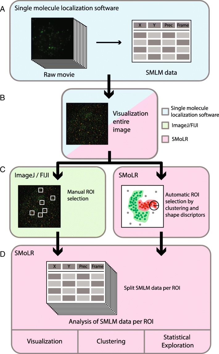
Single-molecule localization microscopy is a super-resolution microscopy technique that allows for nanoscale determination of the localization and organization of proteins in biological samples. For biological interpretation of the data it is essential to extract quantitative information from the super-resolution data sets. Due to the complexity and size of these data sets flexible and user-friendly software is required.

RESULTS

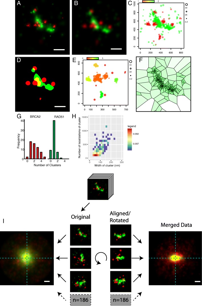
We developed SMoLR (Single Molecule Localization in R): a flexible framework that enables exploration and analysis of single-molecule localization data within the R programming environment. SMoLR is a package aimed at extracting, visualizing and analyzing quantitative information from localization data obtained by single-molecule microscopy. SMoLR is a platform not only to visualize nanoscale subcellular structures but additionally provides means to obtain statistical information about the distribution and localization of molecules within them. This can be done for individual images or SMoLR can be used to analyze a large set of super-resolution images at once. Additionally, we describe a method using SMoLR for image feature-based particle averaging, resulting in identification of common features among nanoscale structures.

CONCLUSIONS

Embedded in the extensive R programming environment, SMoLR allows scientists to study the nanoscale organization of biomolecules in cells by extracting and visualizing quantitative information and hence provides insight in a wide-variety of different biological processes at the single-molecule level.

摘要

背景

单分子定位显微镜是一种超分辨率显微镜技术,可用于确定生物样本中蛋白质的纳米级定位和组织。为了对数据进行生物学解释,必须从超分辨率数据集提取定量信息。由于这些数据集的复杂性和规模,需要灵活且用户友好的软件。

结果

我们开发了 SMoLR(在 R 中进行单分子定位):一个灵活的框架,允许在 R 编程环境中探索和分析单分子定位数据。SMoLR 是一个旨在从单分子显微镜获得的定位数据中提取、可视化和分析定量信息的软件包。SMoLR 不仅是一个可视化纳米级亚细胞结构的平台,还提供了获取有关分子在其中分布和定位的统计信息的方法。可以针对单个图像执行此操作,或者可以使用 SMoLR 一次分析大量超分辨率图像。此外,我们描述了一种使用 SMoLR 进行基于图像特征的粒子平均的方法,从而可以识别纳米级结构之间的常见特征。

结论

嵌入广泛的 R 编程环境中,SMoLR 允许科学家通过提取和可视化定量信息来研究细胞中生物分子的纳米级组织,从而在单细胞水平上深入了解各种不同的生物学过程。

https://cdn.ncbi.nlm.nih.gov/pmc/blobs/1825/6334411/7e521fb7e343/12859_2018_2578_Fig2_HTML.jpg
https://cdn.ncbi.nlm.nih.gov/pmc/blobs/1825/6334411/2403345f5d65/12859_2018_2578_Fig1_HTML.jpg
https://cdn.ncbi.nlm.nih.gov/pmc/blobs/1825/6334411/7e521fb7e343/12859_2018_2578_Fig2_HTML.jpg
https://cdn.ncbi.nlm.nih.gov/pmc/blobs/1825/6334411/2403345f5d65/12859_2018_2578_Fig1_HTML.jpg
https://cdn.ncbi.nlm.nih.gov/pmc/blobs/1825/6334411/7e521fb7e343/12859_2018_2578_Fig2_HTML.jpg

相似文献

1
SMoLR: visualization and analysis of single-molecule localization microscopy data in R.SMoLR:R 中的单分子定位显微镜数据的可视化和分析。
BMC Bioinformatics. 2019 Jan 15;20(1):30. doi: 10.1186/s12859-018-2578-3.
2
Extracting quantitative information from single-molecule super-resolution imaging data with LAMA - LocAlization Microscopy Analyzer.使用 LAMA - LocAlization Microscopy Analyzer 从单分子超分辨率成像数据中提取定量信息。
Sci Rep. 2016 Oct 5;6:34486. doi: 10.1038/srep34486.
3
iSBatch: a batch-processing platform for data analysis and exploration of live-cell single-molecule microscopy images and other hierarchical datasets.iSBatch:一个用于对活细胞单分子显微镜图像和其他分层数据集进行数据分析与探索的批处理平台。
Mol Biosyst. 2015 Oct;11(10):2699-708. doi: 10.1039/c5mb00321k.
4
Super-resolution microscopy with DNA-PAINT.DNA-PAINT 超高分辨率显微镜技术
Nat Protoc. 2017 Jun;12(6):1198-1228. doi: 10.1038/nprot.2017.024. Epub 2017 May 18.
5
Extracting microtubule networks from superresolution single-molecule localization microscopy data.从超分辨率单分子定位显微镜数据中提取微管网络
Mol Biol Cell. 2017 Jan 15;28(2):333-345. doi: 10.1091/mbc.E16-06-0421. Epub 2016 Nov 16.
6
Super-Resolution Fluorescence Microscopy for Single Cell Imaging.超高分辨率荧光显微镜用于单细胞成像。
Adv Exp Med Biol. 2018;1068:59-71. doi: 10.1007/978-981-13-0502-3_6.
7
PALMsiever: a tool to turn raw data into results for single-molecule localization microscopy.PALMsiever:一种将原始数据转化为单分子定位显微镜结果的工具。
Bioinformatics. 2015 Mar 1;31(5):797-8. doi: 10.1093/bioinformatics/btu720. Epub 2014 Oct 31.
8
ThunderSTORM: a comprehensive ImageJ plug-in for PALM and STORM data analysis and super-resolution imaging.ThunderSTORM:一款用于PALM和STORM数据分析及超分辨率成像的综合性ImageJ插件。
Bioinformatics. 2014 Aug 15;30(16):2389-90. doi: 10.1093/bioinformatics/btu202. Epub 2014 Apr 25.
9
Studying the organization of DNA repair by single-cell and single-molecule imaging.通过单细胞和单分子成像研究DNA修复的组织方式。
DNA Repair (Amst). 2014 Aug;20(100):32-40. doi: 10.1016/j.dnarep.2014.02.015. Epub 2014 Mar 12.
10
Localization microscopy coming of age: from concepts to biological impact.定位显微镜崭露头角:从概念到生物影响。
J Cell Sci. 2013 Aug 15;126(Pt 16):3505-13. doi: 10.1242/jcs.123612.

引用本文的文献

1
Distinct mobility patterns of BRCA2 molecules at DNA damage sites.BRCA2 分子在 DNA 损伤部位的独特迁移模式。
Nucleic Acids Res. 2024 Aug 12;52(14):8332-8343. doi: 10.1093/nar/gkae559.
2
Compartmentalization of androgen receptors at endogenous genes in living cells.活细胞内内源基因雄激素受体的区室化。
Nucleic Acids Res. 2023 Nov 10;51(20):10992-11009. doi: 10.1093/nar/gkad803.
3
Three-color dSTORM Imaging and Analysis of Recombination Foci in Mouse Spread Meiotic Nuclei.小鼠减数分裂铺展细胞核中重组位点的三色dSTORM成像与分析

本文引用的文献

1
3DClusterViSu: 3D clustering analysis of super-resolution microscopy data by 3D Voronoi tessellations.3DClusterViSu:通过 3D voronoi 胞腔进行超分辨率显微镜数据的 3D 聚类分析。
Bioinformatics. 2018 Sep 1;34(17):3004-3012. doi: 10.1093/bioinformatics/bty200.
2
Single-molecule localization microscopy reveals molecular transactions during RAD51 filament assembly at cellular DNA damage sites.单分子定位显微镜揭示了细胞 DNA 损伤部位 RAD51 丝组装过程中的分子转位。
Nucleic Acids Res. 2018 Mar 16;46(5):2398-2416. doi: 10.1093/nar/gkx1303.
3
Investigating Focal Adhesion Substructures by Localization Microscopy.
Bio Protoc. 2023 Jul 20;13(14):e4780. doi: 10.21769/BioProtoc.4780.
4
Multi-color dSTORM microscopy in Hormad1-/- spermatocytes reveals alterations in meiotic recombination intermediates and synaptonemal complex structure.多色 dSTORM 显微镜在 Hormad1-/- 精母细胞中显示减数分裂重组中间体和联会复合体结构的改变。
PLoS Genet. 2022 Jul 20;18(7):e1010046. doi: 10.1371/journal.pgen.1010046. eCollection 2022 Jul.
5
Can super-resolution microscopy become a standard characterization technique for materials chemistry?超分辨率显微镜能否成为材料化学的标准表征技术?
Chem Sci. 2021 Dec 1;13(8):2152-2166. doi: 10.1039/d1sc05506b. eCollection 2022 Feb 23.
6
Measuring Nanoscale Distances by Structured Illumination Microscopy and Image Cross-Correlation Spectroscopy (SIM-ICCS).通过结构光照明显微镜和图像互相关光谱学(SIM-ICCS)测量纳米级距离。
Sensors (Basel). 2021 Mar 12;21(6):2010. doi: 10.3390/s21062010.
7
A Review of Super-Resolution Single-Molecule Localization Microscopy Cluster Analysis and Quantification Methods.超分辨率单分子定位显微镜簇分析与量化方法综述
Patterns (N Y). 2020 Jun 12;1(3):100038. doi: 10.1016/j.patter.2020.100038.
8
Super-resolution imaging of RAD51 and DMC1 in DNA repair foci reveals dynamic distribution patterns in meiotic prophase.RAD51 和 DMC1 在 DNA 修复焦点中的超分辨率成像揭示了减数分裂前期的动态分布模式。
PLoS Genet. 2020 Jun 5;16(6):e1008595. doi: 10.1371/journal.pgen.1008595. eCollection 2020 Jun.
9
Redundant and specific roles of cohesin STAG subunits in chromatin looping and transcriptional control.黏连蛋白 STAG 亚基在染色质环化和转录调控中的冗余和特异作用。
Genome Res. 2020 Apr;30(4):515-527. doi: 10.1101/gr.253211.119. Epub 2020 Apr 6.
10
Quantitative Analysis of Nuclear Lamins Imaged by Super-Resolution Light Microscopy.超分辨率荧光显微镜成像的核纤层蛋白的定量分析。
Cells. 2019 Apr 18;8(4):361. doi: 10.3390/cells8040361.
通过定位显微镜研究粘着斑亚结构
Biophys J. 2017 Dec 5;113(11):2508-2518. doi: 10.1016/j.bpj.2017.09.032.
4
Angular reconstitution-based 3D reconstructions of nanomolecular structures from superresolution light-microscopy images.基于角重构成像的超分辨荧光显微图像中纳米分子结构的三维重构。
Proc Natl Acad Sci U S A. 2017 Aug 29;114(35):9273-9278. doi: 10.1073/pnas.1704908114. Epub 2017 Aug 15.
5
Robust nonparametric quantification of clustering density of molecules in single-molecule localization microscopy.单分子定位显微镜中分子聚集密度的稳健非参数定量分析
PLoS One. 2017 Jun 21;12(6):e0179975. doi: 10.1371/journal.pone.0179975. eCollection 2017.
6
Architectural plasticity of human BRCA2-RAD51 complexes in DNA break repair.人类BRCA2-RAD51复合物在DNA断裂修复中的结构可塑性。
Nucleic Acids Res. 2017 May 5;45(8):4507-4518. doi: 10.1093/nar/gkx084.
7
Extracting quantitative information from single-molecule super-resolution imaging data with LAMA - LocAlization Microscopy Analyzer.使用 LAMA - LocAlization Microscopy Analyzer 从单分子超分辨率成像数据中提取定量信息。
Sci Rep. 2016 Oct 5;6:34486. doi: 10.1038/srep34486.
8
SharpViSu: integrated analysis and segmentation of super-resolution microscopy data.SharpViSu:超分辨率显微镜数据的综合分析与分割
Bioinformatics. 2016 Jul 15;32(14):2239-41. doi: 10.1093/bioinformatics/btw123. Epub 2016 Mar 18.
9
ClusterViSu, a method for clustering of protein complexes by Voronoi tessellation in super-resolution microscopy.ClusterViSu,一种在超分辨率显微镜中通过Voronoi镶嵌对蛋白质复合物进行聚类的方法。
Sci Rep. 2016 Apr 12;6:24084. doi: 10.1038/srep24084.
10
Bayesian cluster identification in single-molecule localization microscopy data.贝叶斯聚类在单分子定位显微镜数据中的应用。
Nat Methods. 2015 Nov;12(11):1072-6. doi: 10.1038/nmeth.3612. Epub 2015 Oct 5.