Niehues Stefan M, Denecke Timm, Bassir Christian, Hamm Bernd, Haas Matthias
Klinik für Radiologie, Charité - Universitätsmedizin Berlin, Berlin, Germany.
Acta Radiol Open. 2019 Apr 8;8(4):2058460119836256. doi: 10.1177/2058460119836256. eCollection 2019 Apr.
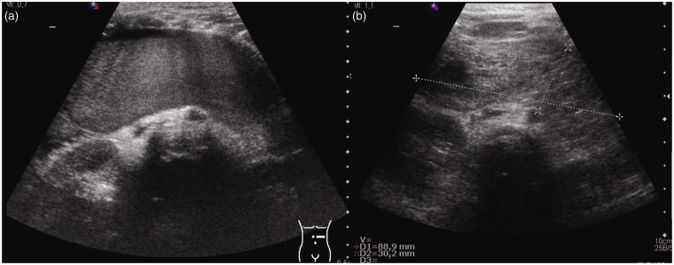
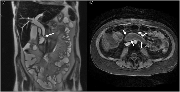
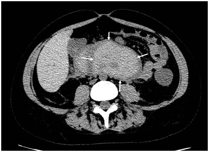
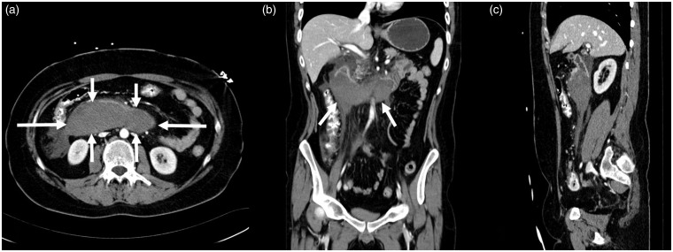
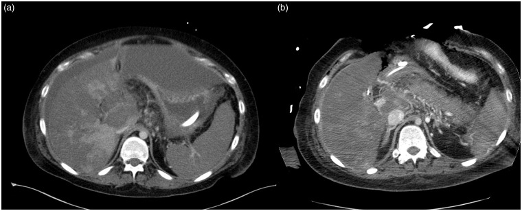
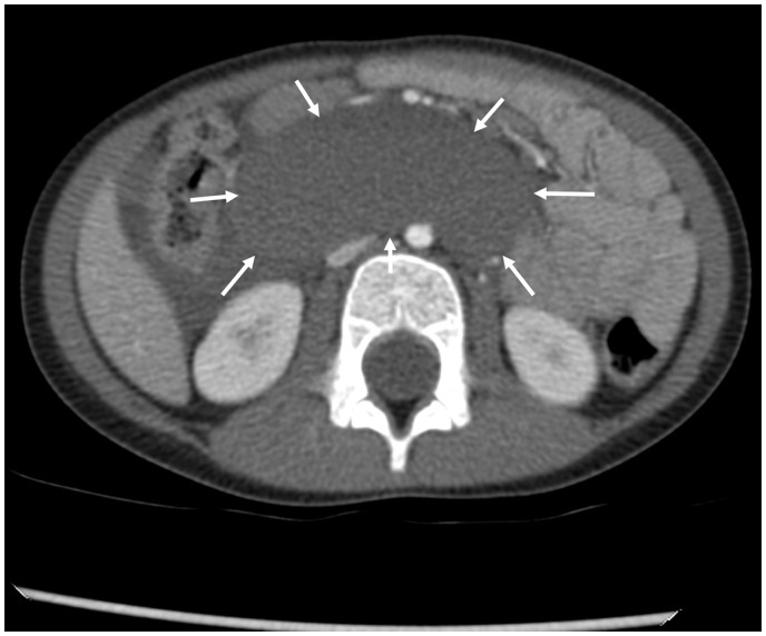
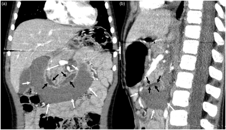
Intramural duodenal hematoma is a rare condition. Different imaging modalities are at hand for diagnosis.
To identify patients with intramural duodenal hematoma and report imaging findings and clinical courses.
Typical imaging patterns using ultrasound, computed tomography, and magnetic resonance imaging were carried out on 10 patients.
The mean patient age was 7.5 years. The average disease duration was 13 months. Clinical signs of improvement were observed within 16 days. Residues were still detectable at long-term follow-up.
For patients with intramural duodenal wall hematoma, diagnosis should be considered early. Typical imaging findings should be known to ensure optimal treatment.
十二指肠壁内血肿是一种罕见病症。有多种不同的成像方式可用于诊断。
识别患有十二指肠壁内血肿的患者,并报告成像结果及临床病程。
对10例患者进行了使用超声、计算机断层扫描和磁共振成像的典型成像检查。
患者平均年龄为7.5岁。平均病程为13个月。在16天内观察到临床改善迹象。在长期随访中仍可检测到残留物。
对于患有十二指肠壁内血肿的患者,应尽早考虑诊断。应了解典型的成像结果以确保最佳治疗。