Chen Cheng, Lv Liang, Hu Yu, Yin Senlin, Zhou Peizhi, Jiang Shu
Department of Neurosurgery, West China Hospital of Sichuan University, Chengdu, Sichuan Province, China.
Medicine (Baltimore). 2019 Apr;98(16):e15334. doi: 10.1097/MD.0000000000015334.
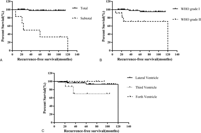
Intraventricular meningiomas are rather rare and only represent a small proportion of all intracranial meningiomas. Data are still limited toward this peculiar entity and surgical resection remains challenging for neurosurgeons. The purpose of present study is to demonstrate clinical features, surgical treatment, and potential risk factors determined long-term prognosis of intraventricular meningiomas.A total of 89 surgically treated and histopathologically confirmed intraventricular meningiomas were identified in our institution from 2008 to 2018. Clinical features, neuroimaging findings, surgical records, and prognosis data were extracted and reviewed retrospectively. Group comparison and recurrence-free survival analysis were performed.Female predominance was well established with a sex radio of 2.1:1. Raised intracranial pressure and decline of visual acuity were 2 chief symptoms that patients generally complained of. Preoperative magnetic resonance imaging (MRI) scans were performed in all patients and there was a trend toward lateral ventricular meningiomas were larger in size than others (P = .07). Superior parietal lobule and temporal approach were widely adopted and lateral/4th ventricular meningiomas were more easily to acquire total tumor resection as compared with 3rd ventricular meningiomas (P = .03). After an average follow-up of 57.3 months, 6 patients experienced recurrence of disease in our series. Individuals with subtotal resection (P < .001) and higher World Health Organization classification (P = .001) were more prone to relapse.Intraventricular meningiomas presented with a wide variety of symptoms. Surgery remained 1st treatment of choice and optimal surgical approach should be planned individually based on preoperative MRI evaluation. Patients underwent subtotal tumor resection and with malignant tumor nature should be carefully monitored during follow-up.
脑室内脑膜瘤相当罕见,仅占所有颅内脑膜瘤的一小部分。针对这一特殊类型肿瘤的数据仍然有限,手术切除对神经外科医生来说仍然具有挑战性。本研究的目的是阐述脑室内脑膜瘤的临床特征、手术治疗以及决定其长期预后的潜在危险因素。
2008年至2018年期间,我们机构共识别出89例经手术治疗且组织病理学确诊的脑室内脑膜瘤。回顾性提取并分析了临床特征、神经影像学检查结果、手术记录及预后数据。进行了组间比较和无复发生存分析。
女性占优势,性别比为2.1:1。颅内压升高和视力下降是患者普遍主诉的两大主要症状。所有患者均进行了术前磁共振成像(MRI)扫描,侧脑室脑膜瘤有比其他部位脑膜瘤更大的趋势(P = 0.07)。广泛采用顶叶上部和颞部入路,与第三脑室脑膜瘤相比,侧脑室/第四脑室脑膜瘤更易实现肿瘤全切(P = 0.03)。平均随访57.3个月后,本系列中有6例患者出现疾病复发。次全切除的患者(P < 0.001)和世界卫生组织分级较高的患者(P = 0.001)更容易复发。
脑室内脑膜瘤表现出多种多样的症状。手术仍然是首选的治疗方法,应根据术前MRI评估为患者个体化制定最佳手术方案。接受次全肿瘤切除且肿瘤性质为恶性的患者在随访期间应密切监测。