• 文献检索
  • 文档翻译
  • 深度研究
  • 学术资讯
  • Suppr Zotero 插件Zotero 插件
  • 邀请有礼
  • 套餐&价格
  • 历史记录
应用&插件
Suppr Zotero 插件Zotero 插件浏览器插件Mac 客户端Windows 客户端微信小程序
定价
高级版会员购买积分包购买API积分包
服务
文献检索文档翻译深度研究API 文档MCP 服务
关于我们
关于 Suppr公司介绍联系我们用户协议隐私条款
关注我们

Suppr 超能文献

核心技术专利:CN118964589B侵权必究
粤ICP备2023148730 号-1Suppr @ 2026

文献检索

告别复杂PubMed语法,用中文像聊天一样搜索,搜遍4000万医学文献。AI智能推荐,让科研检索更轻松。

立即免费搜索

文件翻译

保留排版,准确专业,支持PDF/Word/PPT等文件格式,支持 12+语言互译。

免费翻译文档

深度研究

AI帮你快速写综述,25分钟生成高质量综述,智能提取关键信息,辅助科研写作。

立即免费体验

相似文献

1
Single-Cell Lymphocyte Heterogeneity in Advanced Cutaneous T-cell Lymphoma Skin Tumors.晚期皮肤 T 细胞淋巴瘤皮肤肿瘤中单细胞淋巴细胞异质性。
Clin Cancer Res. 2019 Jul 15;25(14):4443-4454. doi: 10.1158/1078-0432.CCR-19-0148. Epub 2019 Apr 22.
2
Single-Cell Profiling of Cutaneous T-Cell Lymphoma Reveals Underlying Heterogeneity Associated with Disease Progression.单细胞分析揭示皮肤 T 细胞淋巴瘤的潜在异质性与疾病进展相关。
Clin Cancer Res. 2019 May 15;25(10):2996-3005. doi: 10.1158/1078-0432.CCR-18-3309. Epub 2019 Feb 4.
3
Primary T Cells from Cutaneous T-cell Lymphoma Skin Explants Display an Exhausted Immune Checkpoint Profile.原发性皮肤 T 细胞淋巴瘤皮肤外植体中的初始 T 细胞表现出衰竭的免疫检查点特征。
Cancer Immunol Res. 2018 Aug;6(8):900-909. doi: 10.1158/2326-6066.CIR-17-0270. Epub 2018 Jun 12.
4
Lesional gene expression profiling in cutaneous T-cell lymphoma reveals natural clusters associated with disease outcome.皮肤T细胞淋巴瘤的病灶基因表达谱分析揭示了与疾病预后相关的自然聚类。
Blood. 2007 Oct 15;110(8):3015-27. doi: 10.1182/blood-2006-12-061507. Epub 2007 Jul 16.
5
Cutaneous T cell lymphoma: the helping hand of dendritic cells.皮肤T细胞淋巴瘤:树突状细胞的助力
Ann N Y Acad Sci. 2001 Sep;941:1-11.
6
High-scatter T cells: a reliable biomarker for malignant T cells in cutaneous T-cell lymphoma.高散射 T 细胞:皮肤 T 细胞淋巴瘤恶性 T 细胞的可靠生物标志物。
Blood. 2011 Feb 10;117(6):1966-76. doi: 10.1182/blood-2010-05-287664. Epub 2010 Dec 9.
7
Role of high-throughput sequencing in the diagnosis of cutaneous T-cell lymphoma.高通量测序在皮肤 T 细胞淋巴瘤诊断中的作用。
J Clin Pathol. 2018 Sep;71(9):814-820. doi: 10.1136/jclinpath-2018-205004. Epub 2018 Apr 10.
8
Localization of clonal T cells to the epidermis in cutaneous T-cell lymphoma.皮肤T细胞淋巴瘤中克隆性T细胞在表皮的定位。
J Am Acad Dermatol. 1994 Nov;31(5 Pt 1):717-23. doi: 10.1016/s0190-9622(94)70231-4.
9
IL-22, but not IL-17, dominant environment in cutaneous T-cell lymphoma.IL-22,而非 IL-17,在皮肤 T 细胞淋巴瘤中占主导地位。
Clin Cancer Res. 2011 Dec 15;17(24):7529-38. doi: 10.1158/1078-0432.CCR-11-1192. Epub 2011 Nov 2.
10
Single-cell RNA sequencing unveils the communications between malignant T and myeloid cells contributing to tumor growth and immunosuppression in cutaneous T-cell lymphoma.单细胞RNA测序揭示了恶性T细胞与髓样细胞之间的通讯,这种通讯促进了皮肤T细胞淋巴瘤的肿瘤生长和免疫抑制。
Cancer Lett. 2022 Dec 28;551:215972. doi: 10.1016/j.canlet.2022.215972. Epub 2022 Oct 17.

引用本文的文献

1
Revolutionizing tumor diagnosis and treatment: the promise of DNA nanotechnology.革新肿瘤诊断与治疗:DNA纳米技术的前景
Front Immunol. 2025 Aug 29;16:1637539. doi: 10.3389/fimmu.2025.1637539. eCollection 2025.
2
An Update on Single-Cell RNA Sequencing in Illuminating Disease Mechanisms of Cutaneous T-Cell Lymphoma.单细胞RNA测序在揭示皮肤T细胞淋巴瘤疾病机制方面的最新进展
Cancers (Basel). 2025 Sep 5;17(17):2921. doi: 10.3390/cancers17172921.
3
sEV-mediated intercellular transformation from MGAT4A to MGAT4A tumor cells via the HOTAIRM1/miR-196b-5p axis promotes apoptosis resistance in CTCL.小细胞外囊泡(sEV)通过HOTAIRM1/miR-196b-5p轴介导从MGAT4A细胞到MGAT4A肿瘤细胞的细胞间转化,促进蕈样肉芽肿(CTCL)中的细胞凋亡抗性。
Oncogene. 2025 Mar 28. doi: 10.1038/s41388-025-03356-6.
4
CD38 in the pathobiology of cutaneous T-cell lymphoma and the potential for combination therapeutic intervention.CD38在皮肤T细胞淋巴瘤病理生物学中的作用及联合治疗干预的潜力
Leukemia. 2025 May;39(5):1146-1156. doi: 10.1038/s41375-025-02551-4. Epub 2025 Mar 8.
5
The Progression of Mycosis Fungoides During Treatment with Mogamulizumab: A BIO-MUSE Case Study of the Tumor and Immune Response in Peripheral Blood and Tissue.莫加莫珠单抗治疗蕈样肉芽肿的病程:一项关于外周血和组织中肿瘤及免疫反应的BIO-MUSE病例研究
Biomedicines. 2025 Jan 14;13(1):186. doi: 10.3390/biomedicines13010186.
6
Multi-Omic Data Integration Suggests Putative Microbial Drivers of Aetiopathogenesis in Mycosis Fungoides.多组学数据整合提示蕈样肉芽肿发病机制中可能的微生物驱动因素。
Cancers (Basel). 2024 Nov 25;16(23):3947. doi: 10.3390/cancers16233947.
7
CDK9 recruits HUWE1 to degrade RARα and offers therapeutic opportunities for cutaneous T-cell lymphoma.细胞周期蛋白依赖性激酶9招募HUWE1以降解视黄酸受体α,并为皮肤T细胞淋巴瘤提供治疗机会。
Nat Commun. 2024 Dec 5;15(1):10594. doi: 10.1038/s41467-024-54354-3.
8
Integrative epidemiology and immunotranscriptomics uncover a risk and potential mechanism for cutaneous lymphoma unmasking or progression with dupilumab therapy.整合流行病学和免疫转录组学揭示了度普利尤单抗治疗皮肤淋巴瘤暴露或进展的风险及潜在机制。
J Allergy Clin Immunol. 2025 May;155(5):1584-1594. doi: 10.1016/j.jaci.2024.10.028. Epub 2024 Nov 7.
9
RUNX2 as a novel biomarker for early identification of patients progressing to advanced-stage mycosis fungoides.RUNX2作为早期识别进展为晚期蕈样肉芽肿患者的新型生物标志物。
Front Oncol. 2024 Oct 7;14:1421443. doi: 10.3389/fonc.2024.1421443. eCollection 2024.
10
The skin microbiome stratifies patients with cutaneous T cell lymphoma and determines event-free survival.皮肤微生物组将患有皮肤 T 细胞淋巴瘤的患者分层,并确定无事件生存。
NPJ Biofilms Microbiomes. 2024 Aug 29;10(1):74. doi: 10.1038/s41522-024-00542-4.

本文引用的文献

1
Single-Cell Profiling of Cutaneous T-Cell Lymphoma Reveals Underlying Heterogeneity Associated with Disease Progression.单细胞分析揭示皮肤 T 细胞淋巴瘤的潜在异质性与疾病进展相关。
Clin Cancer Res. 2019 May 15;25(10):2996-3005. doi: 10.1158/1078-0432.CCR-18-3309. Epub 2019 Feb 4.
2
The PROCLIPI international registry of early-stage mycosis fungoides identifies substantial diagnostic delay in most patients.PROCLIPI 国际早期蕈样肉芽肿登记处发现,大多数患者存在明显的诊断延迟。
Br J Dermatol. 2019 Aug;181(2):350-357. doi: 10.1111/bjd.17258. Epub 2018 Nov 25.
3
Single-cell heterogeneity in Sézary syndrome.塞扎里综合征中的单细胞异质性。
Blood Adv. 2018 Aug 28;2(16):2115-2126. doi: 10.1182/bloodadvances.2018022608.
4
Cancer cell plasticity: Impact on tumor progression and therapy response.肿瘤细胞的可塑性:对肿瘤进展和治疗反应的影响。
Semin Cancer Biol. 2018 Dec;53:48-58. doi: 10.1016/j.semcancer.2018.08.009. Epub 2018 Aug 18.
5
High-throughput sequencing of the T cell receptor β gene identifies aggressive early-stage mycosis fungoides.高通量测序 T 细胞受体 β 基因可鉴定侵袭性早期蕈样肉芽肿。
Sci Transl Med. 2018 May 9;10(440). doi: 10.1126/scitranslmed.aar5894.
6
Emerging roles of epithelial-mesenchymal transition in hematological malignancies.上皮-间充质转化在血液恶性肿瘤中的新作用。
J Biomed Sci. 2018 Apr 23;25(1):37. doi: 10.1186/s12929-018-0440-6.
7
Squamous Cell Carcinoma Antigen 2 (SCCA2, SERPINB4): An Emerging Biomarker for Skin Inflammatory Diseases.鳞状细胞癌抗原 2(SCCA2,SERPINB4):一种新兴的皮肤炎症性疾病生物标志物。
Int J Mol Sci. 2018 Apr 6;19(4):1102. doi: 10.3390/ijms19041102.
8
Estimation of immune cell content in tumour tissue using single-cell RNA-seq data.利用单细胞 RNA 测序数据估算肿瘤组织中的免疫细胞含量。
Nat Commun. 2017 Dec 11;8(1):2032. doi: 10.1038/s41467-017-02289-3.
9
Translation initiation factors and their relevance in cancer.翻译起始因子及其在癌症中的相关性。
Curr Opin Genet Dev. 2018 Feb;48:82-88. doi: 10.1016/j.gde.2017.11.001. Epub 2017 Nov 16.
10
SFRP2/DPP4 and FMO1/LSP1 Define Major Fibroblast Populations in Human Skin.SFRP2/DPP4 和 FMO1/LSP1 定义了人类皮肤中的主要成纤维细胞群体。
J Invest Dermatol. 2018 Apr;138(4):802-810. doi: 10.1016/j.jid.2017.09.045. Epub 2017 Dec 6.

晚期皮肤 T 细胞淋巴瘤皮肤肿瘤中单细胞淋巴细胞异质性。

Single-Cell Lymphocyte Heterogeneity in Advanced Cutaneous T-cell Lymphoma Skin Tumors.

机构信息

Department of Medicine, Division of Rheumatology and Clinical Immunology, University of Pittsburgh School of Medicine, Pittsburgh, Pennsylvania.

Columbia University Medical Center, New York, New York.

出版信息

Clin Cancer Res. 2019 Jul 15;25(14):4443-4454. doi: 10.1158/1078-0432.CCR-19-0148. Epub 2019 Apr 22.

DOI:10.1158/1078-0432.CCR-19-0148
PMID:31010835
原文链接:https://pmc.ncbi.nlm.nih.gov/articles/PMC6635080/
Abstract

PURPOSE

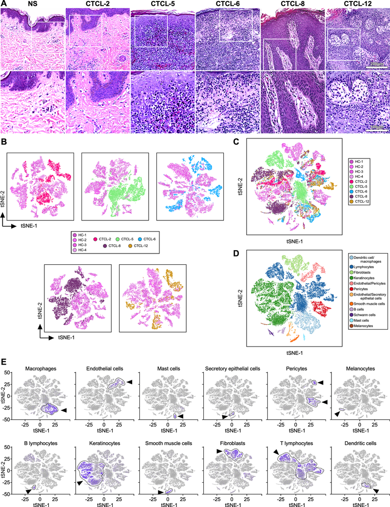
The heterogeneity of tumor cells presents a major challenge to cancer diagnosis and therapy. Cutaneous T-cell lymphomas (CTCL) are a group of T lymphocyte malignancies that primarily affect skin. Lack of highly specific markers for malignant lymphocytes prevents early diagnosis, while only limited treatment options are available for patients with advanced stage CTCL. Droplet-based single-cell transcriptome analysis of CTCL skin biopsies opens avenues for dissecting patient-specific T lymphocyte heterogeneity, providing a basis for identifying specific markers for diagnosis and cure of CTCL.

EXPERIMENTAL DESIGN

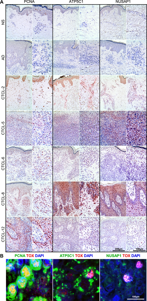
Single-cell RNA-sequencing was performed by Droplet-based sequencing (10X Genomics), focusing on 14,056 CD3 lymphocytes (448 cells from normal and 13,608 cells from CTCL skin samples) from skin biopsies of 5 patients with advanced-stage CTCL and 4 healthy donors. Protein expression of identified genes was validated in advanced stage CTCL skin tumors by immunohistochemistry and confocal immunofluorescence microscopy.

RESULTS

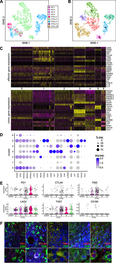
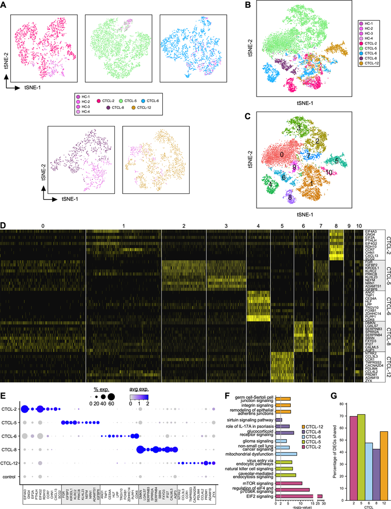
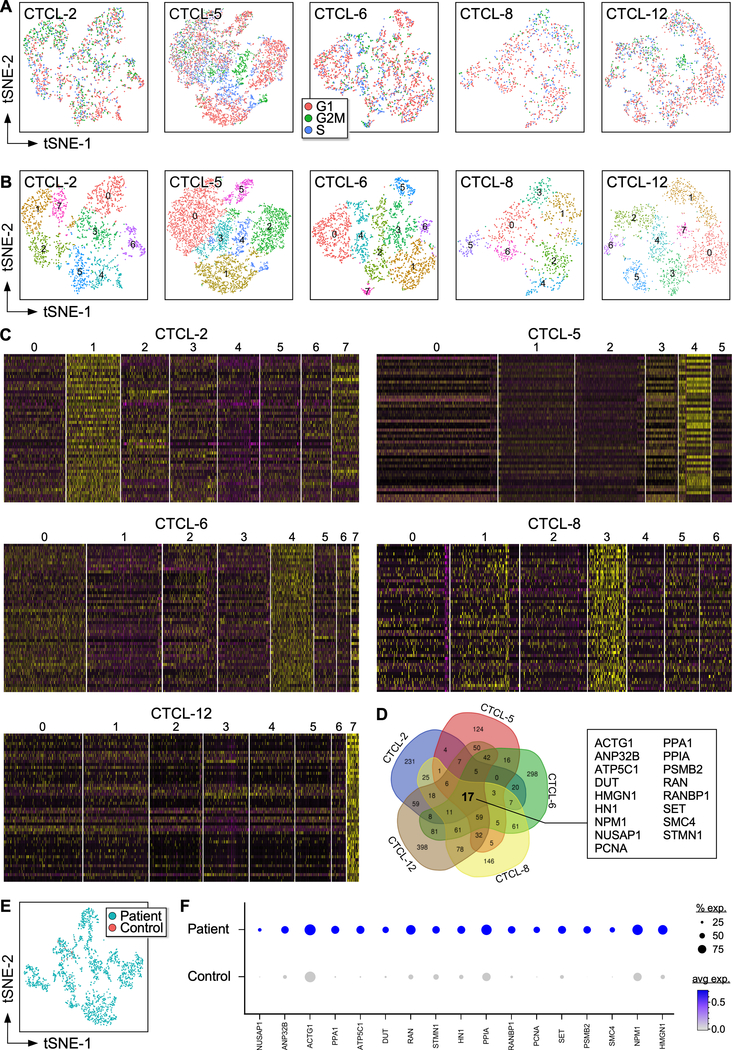
Our analysis revealed a large inter- and intratumor gene expression heterogeneity in the T lymphocyte subset, as well as a common gene expression signature in highly proliferating lymphocytes that was validated in multiple advanced-stage skin tumors. In addition, we established the immunologic state of reactive lymphocytes and found heterogeneity in effector and exhaustion programs across patient samples.

CONCLUSIONS

Single-cell analysis of CTCL skin tumor samples reveals patient-specific landscapes of malignant and reactive lymphocytes within the local microenvironment of each tumor, giving an unprecedented view of lymphocyte heterogeneity and identifying tumor-specific molecular signatures, with important implications for diagnosis and personalized disease treatment.

摘要

目的

肿瘤细胞的异质性对癌症的诊断和治疗构成了重大挑战。皮肤 T 细胞淋巴瘤(CTCL)是一组主要影响皮肤的 T 淋巴细胞恶性肿瘤。恶性淋巴细胞缺乏高度特异性标志物,妨碍了早期诊断,而晚期 CTCL 患者的治疗选择有限。基于液滴的 CTCL 皮肤活检单细胞转录组分析为剖析患者特异性 T 淋巴细胞异质性开辟了途径,为鉴定 CTCL 的诊断和治疗特异性标志物提供了依据。

实验设计

通过基于液滴的测序(10X Genomics)进行单细胞 RNA 测序,重点分析来自 5 名晚期 CTCL 患者和 4 名健康供体皮肤活检的 14,056 个 CD3 淋巴细胞(正常皮肤 448 个细胞,CTCL 皮肤样本 13,608 个细胞)。通过免疫组织化学和共聚焦免疫荧光显微镜验证鉴定基因的蛋白质表达。

结果

我们的分析揭示了 T 淋巴细胞亚群中存在较大的肿瘤内和肿瘤间基因表达异质性,以及在高增殖淋巴细胞中存在共同的基因表达特征,在多个晚期皮肤肿瘤中得到了验证。此外,我们确定了反应性淋巴细胞的免疫状态,并发现了跨患者样本的效应器和衰竭程序的异质性。

结论

对 CTCL 皮肤肿瘤样本的单细胞分析揭示了每个肿瘤局部微环境中恶性和反应性淋巴细胞的患者特异性图谱,为淋巴细胞异质性提供了前所未有的视角,并鉴定了肿瘤特异性分子特征,对诊断和个性化疾病治疗具有重要意义。

https://cdn.ncbi.nlm.nih.gov/pmc/blobs/2709/6635080/40896f7cbffa/nihms-1527465-f0005.jpg
https://cdn.ncbi.nlm.nih.gov/pmc/blobs/2709/6635080/5829ec7c1593/nihms-1527465-f0001.jpg
https://cdn.ncbi.nlm.nih.gov/pmc/blobs/2709/6635080/58bb5f8baa22/nihms-1527465-f0002.jpg
https://cdn.ncbi.nlm.nih.gov/pmc/blobs/2709/6635080/f49ae5953948/nihms-1527465-f0003.jpg
https://cdn.ncbi.nlm.nih.gov/pmc/blobs/2709/6635080/c861f3ff73fb/nihms-1527465-f0004.jpg
https://cdn.ncbi.nlm.nih.gov/pmc/blobs/2709/6635080/40896f7cbffa/nihms-1527465-f0005.jpg