Kong Danli, Yan Yan, He Xiao-Yi, Yang Huihuang, Liang BiYu, Wang Jin, He Yuqing, Ding Yuanlin, Yu Haibing
Department of Epidemiology and Health Statistics, Public Health School of Guangdong Medical University, Dongguan 523808, Guangdong, China.
Academic College of Guangdong Medical University, Dongguan 523808, Guangdong, China.
Biomed Res Int. 2019 Mar 20;2019:8983752. doi: 10.1155/2019/8983752. eCollection 2019.
To observe the effects of resveratrol (Res) on the antioxidative function and estrogen level in an Alzheimer's disease (AD) mouse model.
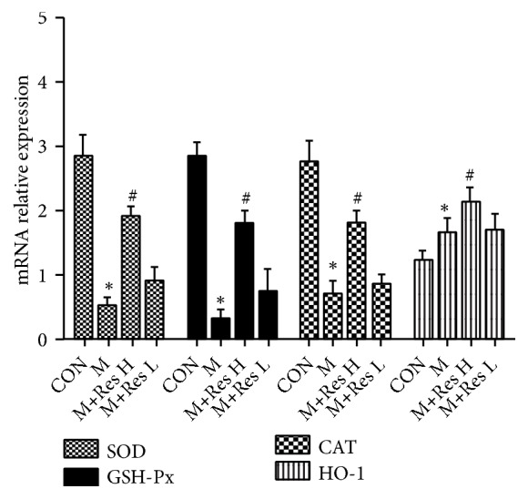
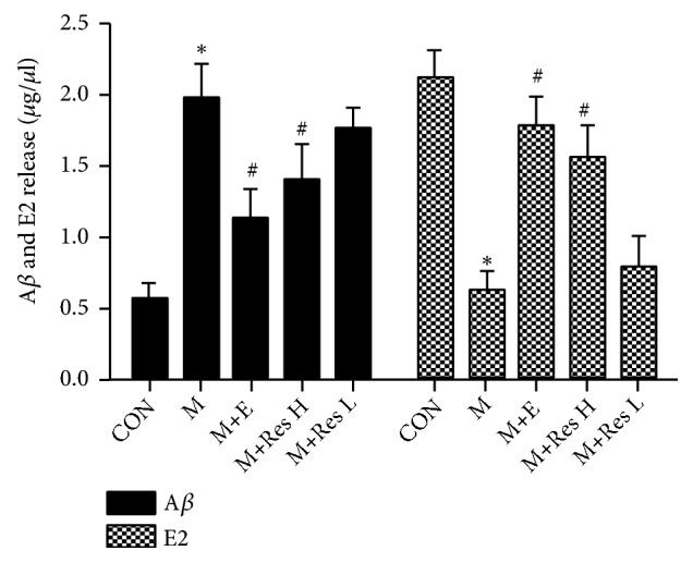
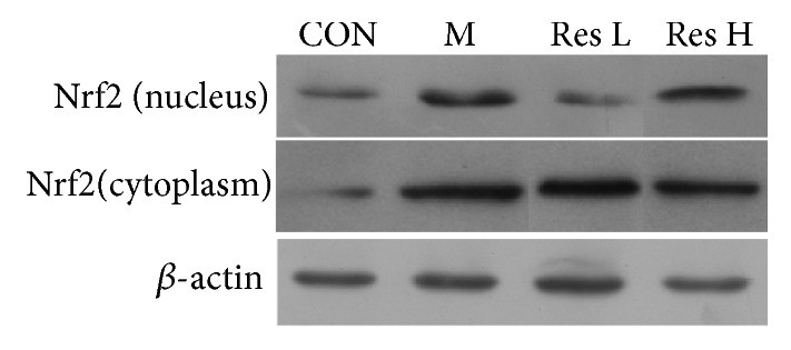
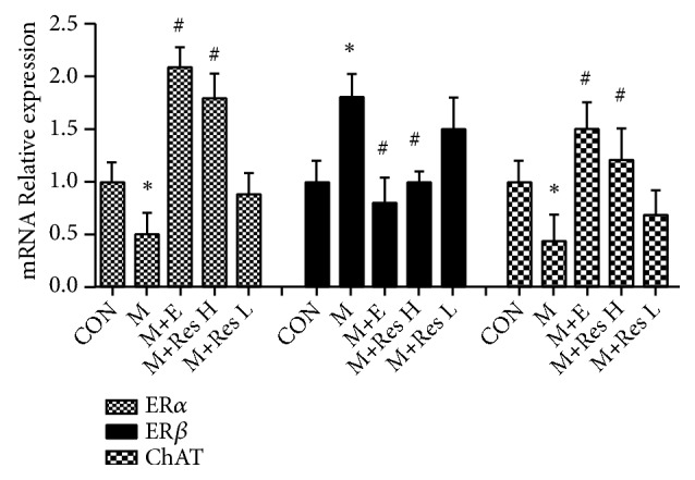
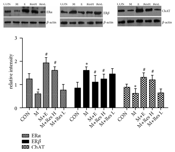
First, we examined the effects of Res on an AD mice model. SAMP8 mice were selected as the model, and normal-aging SAMR1 mice were used as the control group. The model mice were randomly divided into three groups: a model group, high-dose Res group (40mg/kg, intraperitoneal (ip)), and low-dose Res group (20mg/kg, ip). After receiving medication for 15 days, the mice were subjected to the water maze test to assess their spatial discrimination. The spectrophotometric method was used to detect the activity of superoxide dismutase (SOD), glutathione peroxidase (GSH-Px), and catalase (CAT) as well as the malondialdehyde (MDA) content. Quantitative PCR (q-PCR) was used to detect SOD, GSH-Px, CAT, and heme oxygenase-1 (HO-1) mRNA level changes. Western blot analysis detected HO-1 and Nrf2 protein expression. Second, we researched the effect of Res on the estrogen level in the SAMP8 model mice. The model mice were randomly divided into four groups: a model group, estrogen replacement group (0.28 mg/kg, intramuscular (im), estradiol benzoate), high-dose Res group (5 mg/kg, im), and low-dose Res group (2.5 mg/kg, im). The mice were injected, once every three days, for 5 weeks. Q-PCR was used to detect brain tissue mRNA expression changes. Western blot analysis detected ER, ER, and ChAT protein expression. An enzyme-linked immunosorbent assay (ELISA) kit was used to detect the expression of E2 and amyloid protein (A) in brain tissue.
Compared with the control treatment, Res could improve the spatial abilities of the mice to a certain extent and also increase the expression of SOD, GSH-Px, CAT, and HO-1 at the mRNA level (P<0.05). In addition, enhanced SOD, GSH-Px, and CAT activities and HO-1 protein levels and decreased MDA content (P<0.05) were detected in the brain tissue of the Res-treated mice. The cytoplasmic Nrf2 content in the Res-treated mice was also decreased while the nuclear Nrf2 content and the nuclear translation rate of Nrf2 were increased (P<0.05). Res could decrease the expression of ER in the brain tissue at the mRNA and protein levels and the expression of A in the brain tissue at the protein level. Res could also increase the mRNA and protein expression of ER and ChAT and the protein expression of estradiol in the brain tissue.
Res can increase the antioxidant capacity of AD models through the Nrf2/HO-1 signaling pathway. In addition, Res can enhance estrogen levels in an AD model. These findings provide a new idea for the treatment of AD.
观察白藜芦醇(Res)对阿尔茨海默病(AD)小鼠模型抗氧化功能及雌激素水平的影响。
首先,研究Res对AD小鼠模型的作用。选用SAMP8小鼠作为模型,正常衰老的SAMR1小鼠作为对照组。将模型小鼠随机分为三组:模型组、高剂量Res组(40mg/kg,腹腔注射(ip))和低剂量Res组(20mg/kg,ip)。给药15天后,对小鼠进行水迷宫试验以评估其空间辨别能力。采用分光光度法检测超氧化物歧化酶(SOD)、谷胱甘肽过氧化物酶(GSH-Px)和过氧化氢酶(CAT)的活性以及丙二醛(MDA)含量。采用定量聚合酶链反应(q-PCR)检测SOD、GSH-Px、CAT和血红素加氧酶-1(HO-1)mRNA水平的变化。蛋白质免疫印迹分析检测HO-1和Nrf2蛋白表达。其次,研究Res对SAMP8模型小鼠雌激素水平的影响。将模型小鼠随机分为四组:模型组、雌激素替代组(0.28mg/kg,肌肉注射(im),苯甲酸雌二醇)、高剂量Res组(5mg/kg,im)和低剂量Res组(2.5mg/kg,im)。每三天注射一次,共注射5周。采用q-PCR检测脑组织mRNA表达变化。蛋白质免疫印迹分析检测雌激素受体(ER)、ER和胆碱乙酰转移酶(ChAT)蛋白表达。使用酶联免疫吸附测定(ELISA)试剂盒检测脑组织中雌二醇(E2)和淀粉样蛋白(A)的表达。
与对照处理相比,Res能在一定程度上改善小鼠的空间能力,还能增加mRNA水平上SOD、GSH-Px、CAT和HO-1的表达(P<0.05)。此外,在Res处理的小鼠脑组织中检测到SOD、GSH-Px和CAT活性增强,HO-1蛋白水平升高,MDA含量降低(P<0.05)。Res处理的小鼠细胞质中Nrf2含量降低,而细胞核中Nrf2含量和Nrf2的核转位率升高(P<0.05)。Res能降低脑组织中ER在mRNA和蛋白水平的表达以及脑组织中A在蛋白水平的表达。Res还能增加脑组织中ER和ChAT的mRNA和蛋白表达以及雌二醇的蛋白表达。
Res可通过Nrf2/HO-1信号通路提高AD模型的抗氧化能力。此外,Res可提高AD模型中的雌激素水平。这些发现为AD的治疗提供了新思路。