• 文献检索
  • 文档翻译
  • 深度研究
  • 学术资讯
  • Suppr Zotero 插件Zotero 插件
  • 邀请有礼
  • 套餐&价格
  • 历史记录
应用&插件
Suppr Zotero 插件Zotero 插件浏览器插件Mac 客户端Windows 客户端微信小程序
定价
高级版会员购买积分包购买API积分包
服务
文献检索文档翻译深度研究API 文档MCP 服务
关于我们
关于 Suppr公司介绍联系我们用户协议隐私条款
关注我们

Suppr 超能文献

核心技术专利:CN118964589B侵权必究
粤ICP备2023148730 号-1Suppr @ 2026

文献检索

告别复杂PubMed语法,用中文像聊天一样搜索,搜遍4000万医学文献。AI智能推荐,让科研检索更轻松。

立即免费搜索

文件翻译

保留排版,准确专业,支持PDF/Word/PPT等文件格式,支持 12+语言互译。

免费翻译文档

深度研究

AI帮你快速写综述,25分钟生成高质量综述,智能提取关键信息,辅助科研写作。

立即免费体验

使用[F-18]氟雌二醇对雌激素受体阳性乳腺癌进行专用乳腺PET成像的初步经验。

Initial experience of dedicated breast PET imaging of ER+ breast cancers using [F-18]fluoroestradiol.

作者信息

Jones Ella F, Ray Kimberly M, Li Wen, Chien Amy J, Mukhtar Rita A, Esserman Laura J, Franc Benjamin L, Seo Youngho, Pampaloni Miguel H, Joe Bonnie N, Hylton Nola M

机构信息

1Department of Radiology and Biomedical Imaging, University of California, San Francisco, CA USA.

2Helen Diller Family Comprehensive Cancer Center, University of California, San Francisco, CA USA.

出版信息

NPJ Breast Cancer. 2019 Apr 16;5:12. doi: 10.1038/s41523-019-0107-9. eCollection 2019.

DOI:10.1038/s41523-019-0107-9
PMID:31016232
原文链接:https://pmc.ncbi.nlm.nih.gov/articles/PMC6467896/
Abstract

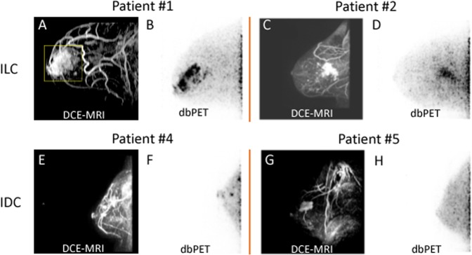
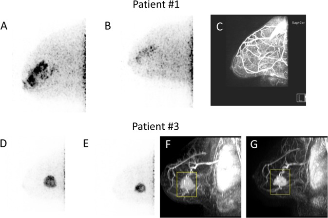
Dedicated breast positron emission tomography (dbPET) is an emerging technology with high sensitivity and spatial resolution that enables detection of sub-centimeter lesions and depiction of intratumoral heterogeneity. In this study, we report our initial experience with dbPET using [F-18]fluoroestradiol (FES) in assessing ER+ primary breast cancers. Six patients with >90% ER+ and HER2- breast cancers were imaged with dbPET and breast MRI. Two patients had ILC, three had IDC, and one had an unknown primary tumor. One ILC patient was treated with letrozole, and another patient with IDC was treated with neoadjuvant chemotherapy without endocrine treatment. In this small cohort, we observed FES uptake in ER+ primary breast tumors with specificity to ER demonstrated in a case with tamoxifen blockade. FES uptake in ILC had a diffused pattern compared to the distinct circumscribed pattern in IDC. In evaluating treatment response, the reduction of SUV was observed with residual disease in an ILC patient treated with letrozole, and an IDC patient treated with chemotherapy. Future study is critical to understand the change in FES SUV after endocrine therapy and to consider other tracer uptake metrics with SUV to describe ER-rich breast cancer. Limitations include variations of FES uptake in different ER+ breast cancer diseases and exclusion of posterior tissues and axillary regions. However, FES-dbPET has a high potential for clinical utility, especially in measuring response to neoadjuvant endocrine treatment. Further development to improve the field of view and studies with a larger cohort of ER+ breast cancer patients are warranted.

摘要

专用乳腺正电子发射断层扫描(dbPET)是一项新兴技术,具有高灵敏度和空间分辨率,能够检测亚厘米级病变并描绘肿瘤内异质性。在本研究中,我们报告了使用[F-18]氟雌二醇(FES)进行dbPET评估雌激素受体阳性(ER+)原发性乳腺癌的初步经验。6例ER+且人表皮生长因子受体2阴性(HER2-)的乳腺癌患者接受了dbPET和乳腺磁共振成像(MRI)检查。2例为小叶原位癌(ILC),3例为浸润性导管癌(IDC),1例原发肿瘤不明。1例ILC患者接受来曲唑治疗,另1例IDC患者接受新辅助化疗但未接受内分泌治疗。在这个小队列中,我们观察到ER+原发性乳腺肿瘤摄取FES,在1例接受他莫昔芬阻断的病例中显示出对ER的特异性。与IDC中明显的边界清晰模式相比,ILC中FES摄取呈弥漫性模式。在评估治疗反应时,接受来曲唑治疗的ILC患者和接受化疗的IDC患者在有残留病灶时观察到标准化摄取值(SUV)降低。未来的研究对于了解内分泌治疗后FES SUV的变化以及考虑使用SUV以外的其他示踪剂摄取指标来描述富含ER的乳腺癌至关重要。局限性包括不同ER+乳腺癌疾病中FES摄取的差异以及后位组织和腋窝区域的排除。然而,FES-dbPET具有很高的临床应用潜力,尤其是在测量对新辅助内分泌治疗的反应方面。有必要进一步改进视野并对更大队列的ER+乳腺癌患者进行研究。

https://cdn.ncbi.nlm.nih.gov/pmc/blobs/4c22/6467896/5e0d7966d2a8/41523_2019_107_Fig2_HTML.jpg
https://cdn.ncbi.nlm.nih.gov/pmc/blobs/4c22/6467896/4f51f022d541/41523_2019_107_Fig1_HTML.jpg
https://cdn.ncbi.nlm.nih.gov/pmc/blobs/4c22/6467896/5e0d7966d2a8/41523_2019_107_Fig2_HTML.jpg
https://cdn.ncbi.nlm.nih.gov/pmc/blobs/4c22/6467896/4f51f022d541/41523_2019_107_Fig1_HTML.jpg
https://cdn.ncbi.nlm.nih.gov/pmc/blobs/4c22/6467896/5e0d7966d2a8/41523_2019_107_Fig2_HTML.jpg

相似文献

1
Initial experience of dedicated breast PET imaging of ER+ breast cancers using [F-18]fluoroestradiol.使用[F-18]氟雌二醇对雌激素受体阳性乳腺癌进行专用乳腺PET成像的初步经验。
NPJ Breast Cancer. 2019 Apr 16;5:12. doi: 10.1038/s41523-019-0107-9. eCollection 2019.
2
A Randomized Feasibility Study of F-Fluoroestradiol PET to Predict Pathologic Response to Neoadjuvant Therapy in Estrogen Receptor-Rich Postmenopausal Breast Cancer.一项关于F-氟雌二醇PET预测富含雌激素受体的绝经后乳腺癌新辅助治疗病理反应的随机可行性研究。
J Nucl Med. 2017 Apr;58(4):563-568. doi: 10.2967/jnumed.116.178368. Epub 2016 Sep 29.
3
Head-to-Head Evaluation of F-FES and F-FDG PET/CT in Metastatic Invasive Lobular Breast Cancer.头对头评估 F-FES 和 F-FDG PET/CT 在转移性浸润性小叶乳腺癌中的应用。
J Nucl Med. 2021 Mar;62(3):326-331. doi: 10.2967/jnumed.120.247882. Epub 2020 Jul 17.
4
The Assessment of Estrogen Receptor Status and Its Intratumoral Heterogeneity in Patients With Breast Cancer by Using 18F-Fluoroestradiol PET/CT.应用 18F-氟雌二醇 PET/CT 评估乳腺癌患者的雌激素受体状态及其肿瘤内异质性。
Clin Nucl Med. 2017 Jun;42(6):421-427. doi: 10.1097/RLU.0000000000001587.
5
Phase II trial of neoadjuvant letrozole and lapatinib in Asian postmenopausal women with estrogen receptor (ER) and human epidermal growth factor receptor 2 (HER2)-positive breast cancer [Neo-ALL-IN]: Highlighting the TILs, ER expressional change after neoadjuvant treatment, and FES-PET as potential significant biomarkers.亚洲绝经后雌激素受体(ER)和人表皮生长因子受体2(HER2)阳性乳腺癌患者新辅助来曲唑和拉帕替尼治疗的II期试验[Neo-ALL-IN]:强调肿瘤浸润淋巴细胞、新辅助治疗后ER表达变化以及FES-PET作为潜在的重要生物标志物
Cancer Chemother Pharmacol. 2016 Oct;78(4):685-95. doi: 10.1007/s00280-016-3107-6. Epub 2016 Aug 4.
6
Can F-Fluoroestradiol Positron Emission Tomography Become a New Imaging Standard in the Estrogen Receptor-positive Breast Cancer Patient: A Prospective Comparative Study with F-Fluorodeoxyglucose Positron Emission Tomography?氟-雌二醇正电子发射断层扫描能否成为雌激素受体阳性乳腺癌患者的新成像标准:与氟-脱氧葡萄糖正电子发射断层扫描的前瞻性对比研究?
World J Nucl Med. 2017 Apr-Jun;16(2):133-139. doi: 10.4103/1450-1147.203071.
7
The Application of 18 F-FES PET in Clinical Cancer Care : A Systematic Review.18 F-FES PET 在临床癌症治疗中的应用:系统评价。
Clin Nucl Med. 2023 Sep 1;48(9):785-795. doi: 10.1097/RLU.0000000000004760. Epub 2023 Jul 22.
8
Can fluorine-18 fluoroestradiol positron emission tomography-computed tomography demonstrate the heterogeneity of breast cancer in vivo?氟-18 氟雌二醇正电子发射断层扫描-计算机断层扫描能在体内显示乳腺癌的异质性吗?
Clin Breast Cancer. 2013 Oct;13(5):359-63. doi: 10.1016/j.clbc.2013.02.012. Epub 2013 Jun 17.
9
The Predictive Value of Early Changes in F-Fluoroestradiol Positron Emission Tomography/Computed Tomography During Fulvestrant 500 mg Therapy in Patients with Estrogen Receptor-Positive Metastatic Breast Cancer.氟[18F]雌二醇正电子发射断层扫描/计算机断层扫描在氟维司群 500mg 治疗雌激素受体阳性转移性乳腺癌患者中早期变化的预测价值。
Oncologist. 2020 Nov;25(11):927-936. doi: 10.1634/theoncologist.2019-0561. Epub 2020 Apr 28.
10
Intratumoral heterogeneity on dedicated breast positron emission tomography predicts malignancy grade of breast cancer.专用乳腺正电子发射断层扫描的肿瘤内异质性可预测乳腺癌的恶性程度。
Breast Cancer Res Treat. 2018 Sep;171(2):315-323. doi: 10.1007/s10549-018-4791-1. Epub 2018 May 19.

引用本文的文献

1
Positive Margin Rates After Breast-Conserving Surgery by Histologic Subtype: A Systematic Review and Meta-analysis Evaluating the Impact of Oncoplastic Surgery.保乳手术后不同组织学亚型的切缘阳性率:一项评估肿瘤整形手术影响的系统评价和荟萃分析
Ann Surg Oncol. 2025 Apr 24. doi: 10.1245/s10434-025-17329-2.
2
Innovations in Positron Emission Tomography and State of the Art in the Evaluation of Breast Cancer Treatment Response.正电子发射断层扫描的创新与乳腺癌治疗反应评估的最新技术水平
J Clin Med. 2023 Dec 27;13(1):154. doi: 10.3390/jcm13010154.
3
Multiparametric Approach to Breast Cancer With Emphasis on Magnetic Resonance Imaging in the Era of Personalized Breast Cancer Treatment.

本文引用的文献

1
TAILORing Adjuvant Systemic Therapy for Breast Cancer.乳腺癌辅助全身治疗的个体化定制
N Engl J Med. 2018 Jul 12;379(2):191-192. doi: 10.1056/NEJMe1806329.
2
Improved visualization of breast tissue on a dedicated breast PET system through ergonomic redesign of the imaging table.通过对成像台进行人体工程学重新设计,在专用乳腺PET系统上改善乳腺组织的可视化。
EJNMMI Res. 2017 Dec 19;7(1):100. doi: 10.1186/s13550-017-0351-7.
3
Dedicated Breast Positron Emission Tomography for the Evaluation of Early Response to Neoadjuvant Chemotherapy in Breast Cancer.
多参数方法在乳腺癌诊治中的应用——个体化乳腺癌治疗时代的磁共振成像重点
Invest Radiol. 2024 Jan 1;59(1):26-37. doi: 10.1097/RLI.0000000000001044. Epub 2023 Nov 22.
4
Advances in Breast PET Instrumentation.乳腺正电子发射断层扫描仪器的进展。
PET Clin. 2024 Jan;19(1):37-47. doi: 10.1016/j.cpet.2023.09.001.
5
[Breast-like cancer of unknown primary : Implications for radiological diagnostics].[原发灶不明的乳腺样癌:对放射诊断的意义]
Radiologie (Heidelb). 2023 May;63(5):366-370. doi: 10.1007/s00117-023-01136-5. Epub 2023 Mar 28.
6
Molecular Imaging for Estrogen Receptor-Positive Breast Cancer: Clinical Applications of Whole Body and Dedicated Breast Positron Emission Tomography.用于雌激素受体阳性乳腺癌的分子成像:全身和专用乳腺正电子发射断层扫描的临床应用。
Surg Oncol Clin N Am. 2022 Oct;31(4):569-579. doi: 10.1016/j.soc.2022.06.001.
7
PET-CT in Clinical Adult Oncology: II. Primary Thoracic and Breast Malignancies.PET-CT在临床成人肿瘤学中的应用:II. 原发性胸部和乳腺恶性肿瘤
Cancers (Basel). 2022 May 29;14(11):2689. doi: 10.3390/cancers14112689.
8
Examining the evolving utility of FDG-PET/CT in breast cancer recurrence.探讨氟代脱氧葡萄糖正电子发射断层扫描/计算机断层扫描(FDG-PET/CT)在乳腺癌复发中的应用进展。
Transl Cancer Res. 2020 Jan;9(Suppl 1):S116-S121. doi: 10.21037/tcr.2019.07.07.
9
Utility of [18F]-fluoroestradiol (FES) PET/CT with dedicated brain acquisition in differentiating brain metastases from posttreatment change in estrogen receptor-positive breast cancer.[18F] - 氟雌二醇(FES)PET/CT联合专用脑部采集在鉴别雌激素受体阳性乳腺癌脑转移与治疗后改变中的应用价值。
Neurooncol Adv. 2021 Nov 30;3(1):vdab178. doi: 10.1093/noajnl/vdab178. eCollection 2021 Jan-Dec.
10
Evaluation of primary breast cancers using dedicated breast PET and whole-body PET.使用专用乳腺 PET 和全身 PET 对原发性乳腺癌进行评估。
Sci Rep. 2020 Dec 14;10(1):21930. doi: 10.1038/s41598-020-78865-3.
专用乳腺正电子发射断层扫描用于评估乳腺癌新辅助化疗的早期反应
Clin Breast Cancer. 2017 Jun;17(3):e155-e159. doi: 10.1016/j.clbc.2016.12.008. Epub 2016 Dec 29.
4
A Randomized Feasibility Study of F-Fluoroestradiol PET to Predict Pathologic Response to Neoadjuvant Therapy in Estrogen Receptor-Rich Postmenopausal Breast Cancer.一项关于F-氟雌二醇PET预测富含雌激素受体的绝经后乳腺癌新辅助治疗病理反应的随机可行性研究。
J Nucl Med. 2017 Apr;58(4):563-568. doi: 10.2967/jnumed.116.178368. Epub 2016 Sep 29.
5
Evaluation of a Hanging-Breast PET System for Primary Tumor Visualization in Patients With Stage I-III Breast Cancer: Comparison With Standard PET/CT.用于I-III期乳腺癌患者原发肿瘤可视化的悬吊式乳房PET系统评估:与标准PET/CT的比较
AJR Am J Roentgenol. 2016 Jun;206(6):1307-14. doi: 10.2214/AJR.15.15371. Epub 2016 Apr 8.
6
Role of Optimal Quantification of FDG PET Imaging in the Clinical Practice of Radiology.正电子发射断层扫描(PET)成像中氟代脱氧葡萄糖(FDG)的最佳量化在放射学临床实践中的作用。
Radiographics. 2016 Mar-Apr;36(2):481-96. doi: 10.1148/rg.2016150102.
7
Discordant assessment of tumor biomarkers by histopathological and molecular assays in the EORTC randomized controlled 10041/BIG 03-04 MINDACT trial breast cancer : Intratumoral heterogeneity and DCIS or normal tissue components are unlikely to be the cause of discordance.在欧洲癌症研究与治疗组织(EORTC)随机对照10041/BIG 03-04 MINDACT试验乳腺癌中,通过组织病理学和分子检测对肿瘤生物标志物进行的不一致评估:瘤内异质性以及导管原位癌或正常组织成分不太可能是不一致的原因。
Breast Cancer Res Treat. 2016 Feb;155(3):463-9. doi: 10.1007/s10549-016-3690-6. Epub 2016 Jan 28.
8
Prospective Validation of a 21-Gene Expression Assay in Breast Cancer.21基因表达检测法在乳腺癌中的前瞻性验证
N Engl J Med. 2015 Nov 19;373(21):2005-14. doi: 10.1056/NEJMoa1510764. Epub 2015 Sep 27.
9
Lobular breast cancer series: imaging.小叶性乳腺癌系列:影像学
Breast Cancer Res. 2015 Jul 11;17(1):94. doi: 10.1186/s13058-015-0605-0.
10
Is the standard uptake value (SUV) appropriate for quantification in clinical PET imaging? - Variability induced by different SUV measurements and varying reconstruction methods.标准摄取值(SUV)是否适用于临床PET成像的定量分析?——不同SUV测量方法和不同重建方法引起的变异性。
Eur J Radiol. 2015 Jan;84(1):158-162. doi: 10.1016/j.ejrad.2014.10.018. Epub 2014 Nov 1.