Department of Oral Cell Biology, Academic Centre for Dentistry Amsterdam (ACTA), University of Amsterdam and Vrije Universiteit Amsterdam, Amsterdam Movement Sciences, Amsterdam, The Netherlands.
Department of Oral and Maxillofacial Surgery, Amsterdam University Medical Centers (Amsterdam UMC)/ACTA, location VUmc, Amsterdam Movement Sciences, Amsterdam, The Netherlands.
J Cell Physiol. 2019 Nov;234(11):20520-20532. doi: 10.1002/jcp.28652. Epub 2019 Apr 23.
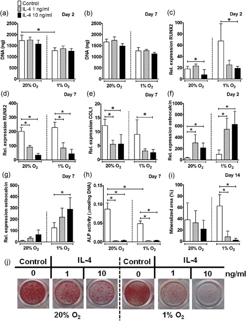
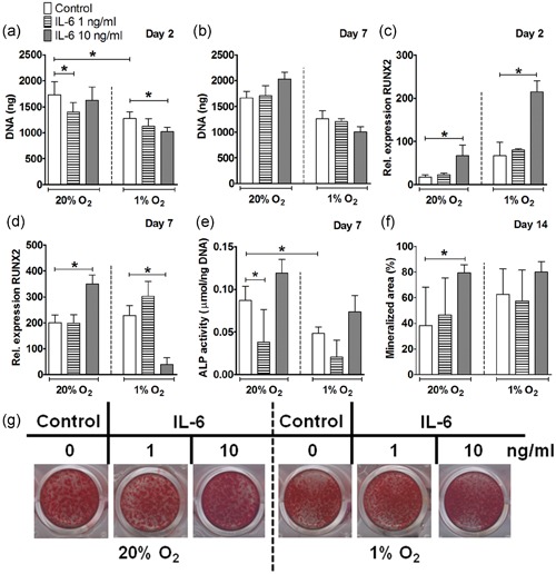
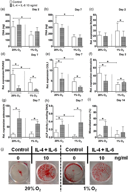
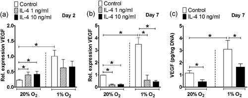
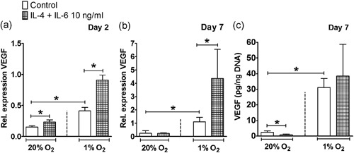
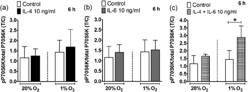
Fracture repair is characterized by cytokine production and hypoxia. To better predict cytokine modulation of mesenchymal stem cell (MSC)-aided bone healing, we investigated whether interleukin 4 (IL-4), IL-6, and their combination, affect osteogenic differentiation, vascular endothelial growth factor (VEGF) production, and/or mammalian target of rapamycin complex 1 (mTORC1) activation by MSCs under normoxia or hypoxia. Human adipose stem cells (hASCs) were cultured with IL-4, IL-6, or their combination for 3 days under normoxia (20% O ) or hypoxia (1% O ), followed by 11 days without cytokines under normoxia or hypoxia. Hypoxia did not alter IL-4 or IL-6-modulated gene or protein expression by hASCs. IL-4 alone decreased runt-related transcription factor 2 (RUNX2) and collagen type 1 (COL1) gene expression, alkaline phosphatase (ALP) activity, and VEGF protein production by hASCs under normoxia and hypoxia, and decreased mineralization of hASCs under hypoxia. In contrast, IL-6 increased mineralization of hASCs under normoxia, and enhanced RUNX2 gene expression under normoxia and hypoxia. Neither IL-4 nor IL-6 affected phosphorylation of the mTORC1 effector protein P70S6K. IL-4 combined with IL-6 diminished the inhibitory effect of IL-4 on ALP activity, bone nodule formation, and VEGF production, and decreased RUNX2 and COL1 expression, similar to IL-4 alone, under normoxia and hypoxia. In conclusion, IL-4 alone, but not in combination with IL-6, inhibits osteogenic differentiation and angiogenic stimulation potential of hASCs under normoxia and hypoxia, likely through pathways other than mTORC1. These results indicate that cytokines may differentially affect bone healing and regeneration when applied in isolation or in combination.
骨折修复的特征在于细胞因子的产生和缺氧。为了更好地预测细胞因子对间充质干细胞(MSC)辅助骨愈合的调节作用,我们研究了白细胞介素 4(IL-4)、白细胞介素 6(IL-6)及其组合是否会影响 MSC 在常氧或低氧条件下的成骨分化、血管内皮生长因子(VEGF)产生和/或哺乳动物雷帕霉素靶蛋白复合物 1(mTORC1)的激活。将人脂肪干细胞(hASCs)在常氧(20% O )或低氧(1% O )下用 IL-4、IL-6 或其组合培养 3 天,然后在常氧或低氧下无细胞因子培养 11 天。低氧不会改变 hASCs 中 IL-4 或 IL-6 调节的基因或蛋白表达。IL-4 单独降低 hASCs 在常氧和低氧下 runt 相关转录因子 2(RUNX2)和胶原蛋白 1(COL1)基因表达、碱性磷酸酶(ALP)活性和 VEGF 蛋白产生,并降低 hASCs 在低氧下的矿化。相比之下,IL-6 增加 hASCs 在常氧下的矿化,并增强 hASCs 在常氧和低氧下的 RUNX2 基因表达。IL-4 和 IL-6 均不影响 mTORC1 效应蛋白 P70S6K 的磷酸化。IL-4 与 IL-6 联合使用可降低 IL-4 对 ALP 活性、骨结节形成和 VEGF 产生的抑制作用,并降低 RUNX2 和 COL1 表达,在常氧和低氧下与单独使用 IL-4 相似。总之,IL-4 单独,而不是与 IL-6 联合使用,可抑制 hASCs 在常氧和低氧下的成骨分化和血管生成刺激潜力,可能通过 mTORC1 以外的途径。这些结果表明,细胞因子在单独或联合使用时可能会对骨愈合和再生产生不同的影响。