Dos Santos Teresa Cristina Sarmet, Setúbal Sérgio, Dos Santos Alair Augusto Sarmet Moreira Damas, Boechat Marcia, Cardoso Claudete Aparecida Araújo
Universidade Federal Fluminense (UFF) - Hospital Universitário Antônio Pedro (HUAP), Niterói, RJ, Brazil.
Instituto Nacional de Saúde da Mulher, da Criança e do Adolescente Fernandes Figueira (IFF/Fiocruz), Rio de Janeiro, RJ, Brazil.
Radiol Bras. 2019 Mar-Apr;52(2):71-77. doi: 10.1590/0100-3984.2018.0025.
To describe the chest computed tomography (CT) findings in immunocompetent children under 36 months of age with pulmonary tuberculosis.
This was a descriptive case series conducted in the city of Rio de Janeiro, Brazil, between January 2004 and July 2013, involving 20 young children who underwent CT after undergoing chest X-rays that did not provide a definitive diagnosis.
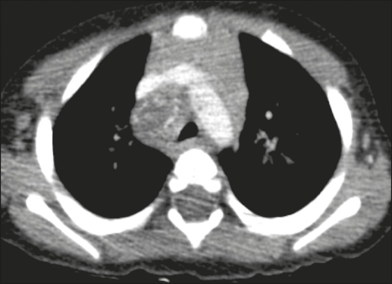
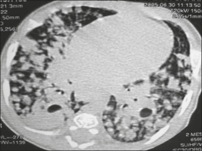
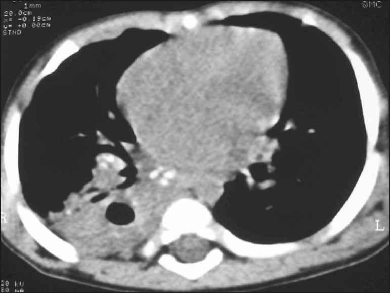
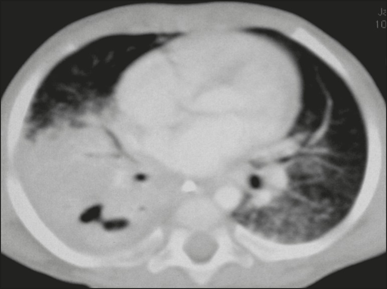
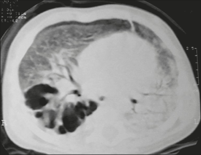
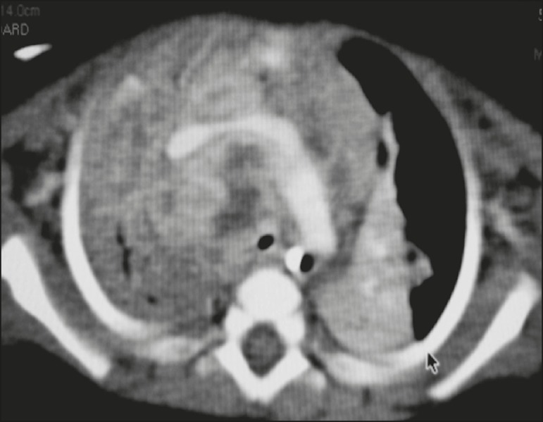
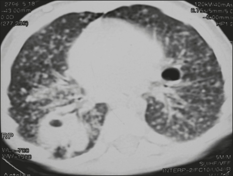
All of the participants had lymph node enlargement and consolidations. In 15 cases (75%), the consolidations were accompanied by atelectasis. Pulmonary cavitation was seen in 10 cases (50%), and cavitation within consolidations was seen in 7 (35%). The areas of cavitation and parenchymal destruction were not seen on conventional chest X-rays.
The radiological presentation of pulmonary tuberculosis in young children differs from that described in older children and adults. CT is an effective method for the early diagnosis of pulmonary tuberculosis in immunocompetent infants, allowing the rapid institution of specific treatment, which is crucial for halting disease progression, as well as for preventing local and systemic complications.
描述36个月以下免疫功能正常的儿童肺结核的胸部计算机断层扫描(CT)表现。
这是一项描述性病例系列研究,于2004年1月至2013年7月在巴西里约热内卢市进行,纳入20名幼儿,这些幼儿在胸部X线检查未能明确诊断后接受了CT检查。
所有参与者均有淋巴结肿大和实变。15例(75%)实变伴有肺不张。10例(50%)可见肺空洞,7例(35%)可见实变内空洞。常规胸部X线检查未见空洞和实质破坏区域。
幼儿肺结核的影像学表现与大龄儿童和成人不同。CT是免疫功能正常婴儿肺结核早期诊断的有效方法,可迅速开始特异性治疗,这对于阻止疾病进展以及预防局部和全身并发症至关重要。