Cappelli Carlo, Pirola Ilenia, Gandossi Elena, Marini Fiorella, Cristiano Alessandra, Casella Claudio, Lombardi Davide, Agosti Barbara, Ferlin Alberto, Castellano Maurizio
Department of Clinical and Experimental Sciences, SSD Medicina ad indirizzo Endocrino-metabolico, University of Brescia, ASST Spedali Civili di Brescia, 25123 Brescia, Italy.
Department of Molecular and Translational Medicine, 3rd Division of General Surgery, University of Brescia, ASST Spedali Civili di Brescia, 25123 Brescia, Italy.
Int J Endocrinol. 2019 Mar 25;2019:7874890. doi: 10.1155/2019/7874890. eCollection 2019.
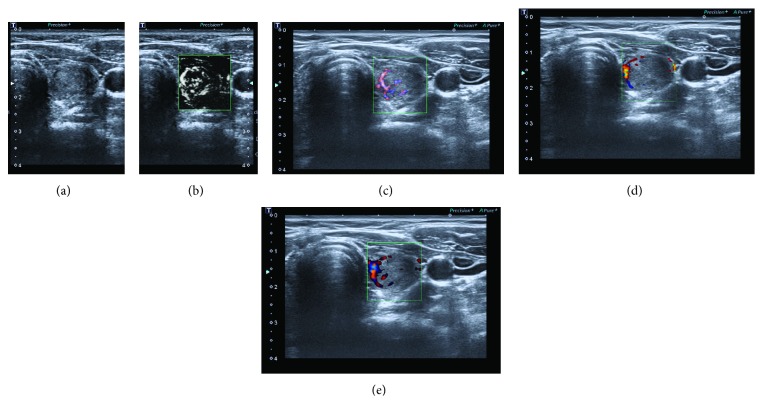
Toshiba Medical System has developed a new Doppler technique [Superb Microvascular Imaging (SMI)] that has improved microvascular flow imaging. SMI depicts perinodular and intranodular thyroid microvascular flow in higher detail compared to standard colour Doppler (CD) and power Doppler (PD) imaging.
Assess the nodular microvascular architecture by SMI compared to CD and PD features in a series of thyroid nodules submitted to fine needle aspiration cytology, in order to evaluate the potential of SMI in detecting thyroid cancer.
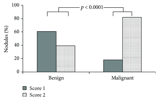
From April 2016 to July 2017, 254 patients with thyroid nodules, evaluated as at high risk for malignancy in agreement with AACE/ACE/AME guidelines, were submitted to cytology. All nodules were previously submitted to ultrasound grayscale, CD, PD, and SMI evaluation. Benign and malignant nodules were stratified in accordance to the number of vessels visualised by SMI: score 1 with a maximum of two blood vessels and score 2 with three or more vessels.
Score 1 was found in 59.6% of benign nodules and in 17.9% of malignant nodules, whereas score 2 was found in 40.4% and in 82.1%, respectively (sensitivity 81.7%; specificity 60.5%, < 0.001). Variables significantly associated with malignancy in the univariate analysis were gender (OR, 0.18; 95% CI, 0.08-0.37; < 0.001), vascularity (OR, 1.91; 95% CI, 1.65-3.89; < 0.001), and SMI (OR, 6.72; 95% CI, 3.89-11.59; < 0.001); multivariate logistic model confirmed SMI score 2 as an independent risk factor for malignancy (OR, 6.99; 95% CI, 3.46-12.09; < 0.001).
This prospective pilot study showed that SMI can depict intranodular flow in higher detail compared to CDI and PDI, thus improving thyroid cancer detection.
东芝医疗系统公司开发了一种新的多普勒技术[超微血管成像(SMI)],该技术改善了微血管血流成像。与标准彩色多普勒(CD)和能量多普勒(PD)成像相比,SMI能更详细地描绘甲状腺结节周围和结节内的微血管血流。
在一系列接受细针穿刺细胞学检查的甲状腺结节中,将SMI与CD和PD特征进行比较,评估结节的微血管结构,以评估SMI在检测甲状腺癌方面的潜力。
2016年4月至2017年7月,254例根据美国临床内分泌医师协会(AACE)/美国内分泌学会(ACE)/美国医学内分泌学会(AME)指南被评估为恶性风险高的甲状腺结节患者接受了细胞学检查。所有结节之前均接受了超声灰阶、CD、PD和SMI评估。根据SMI显示的血管数量对良性和恶性结节进行分层:1分表示最多有两条血管,2分表示有三条或更多血管。
1分在59.6%的良性结节和17.9%的恶性结节中出现,而2分分别在40.4%和82.1%的结节中出现(敏感性81.7%;特异性60.5%,P<0.001)。单因素分析中与恶性肿瘤显著相关的变量为性别(比值比[OR],0.18;95%置信区间[CI],0.08 - 0.37;P<0.001)、血管分布(OR,1.91;95%CI,1.65 - 3.89;P<0.001)和SMI(OR,6.72;95%CI,3.89 - 11.59;P<0.001);多因素逻辑模型证实SMI 2分为恶性肿瘤的独立危险因素(OR,6.99;95%CI,3.46 - 12.09;P<0.001)。
这项前瞻性初步研究表明,与CDI和PDI相比,SMI能更详细地描绘结节内血流,从而提高甲状腺癌的检测率。