• 文献检索
  • 文档翻译
  • 深度研究
  • 学术资讯
  • Suppr Zotero 插件Zotero 插件
  • 邀请有礼
  • 套餐&价格
  • 历史记录
应用&插件
Suppr Zotero 插件Zotero 插件浏览器插件Mac 客户端Windows 客户端微信小程序
定价
高级版会员购买积分包购买API积分包
服务
文献检索文档翻译深度研究API 文档MCP 服务
关于我们
关于 Suppr公司介绍联系我们用户协议隐私条款
关注我们

Suppr 超能文献

核心技术专利:CN118964589B侵权必究
粤ICP备2023148730 号-1Suppr @ 2026

文献检索

告别复杂PubMed语法,用中文像聊天一样搜索,搜遍4000万医学文献。AI智能推荐,让科研检索更轻松。

立即免费搜索

文件翻译

保留排版,准确专业,支持PDF/Word/PPT等文件格式,支持 12+语言互译。

免费翻译文档

深度研究

AI帮你快速写综述,25分钟生成高质量综述,智能提取关键信息,辅助科研写作。

立即免费体验

腹部结节病的超声成像:最新进展

Ultrasound imaging of abdominal sarcoidosis: State of the art.

作者信息

Tana Claudio, Schiavone Cosima, Ticinesi Andrea, Ricci Fabrizio, Giamberardino Maria Adele, Cipollone Francesco, Silingardi Mauro, Meschi Tiziana, Dietrich Christoph F

机构信息

Internal Medicine and Critical Subacute Care Unit, Medicine Geriatric-Rehabilitation Department, and Department of Medicine and Surgery, University-Hospital of Parma, Parma 43126, Italy.

Department of Internistic Ultrasound, "G. D'Annunzio" University of Chieti, Chieti 66100, Italy.

出版信息

World J Clin Cases. 2019 Apr 6;7(7):809-818. doi: 10.12998/wjcc.v7.i7.809.

DOI:10.12998/wjcc.v7.i7.809
PMID:31024952
原文链接:https://pmc.ncbi.nlm.nih.gov/articles/PMC6473121/
Abstract

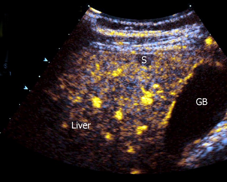
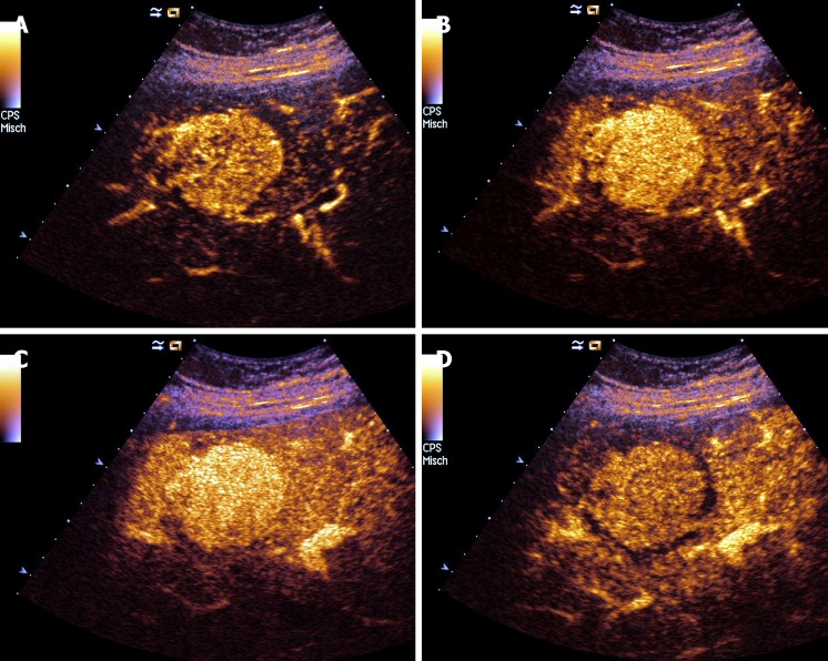
Since it has been recognized that sarcoidosis (SA) is not an exclusive disorder of the lungs but can also affect other organs such as the liver and spleen, efforts have been made to define specific imaging criteria for the diagnosis of the single organ involvement, and the concept has been reinforced that the exclusion of alternative causes is important to achieve the correct diagnosis. Ultrasound (US) is a useful tool to evaluate patients with suspected abdominal SA, such as of the liver, spleen, kidney, pancreas and other organs, showing findings such as organomegaly, focal lesions and lymphadenopathy. While the diagnosis of abdominal SA is more predictable in the case of involvement of other organs (., lungs), the problem is more complex in the case of isolated abdominal SA. The recent use of contrast-enhanced ultrasound and endoscopic ultrasound elastography has provided additional information about the enhancement patterns and tissue rigidity in abdominal SA. Here we critically review the role of US in abdominal SA, reporting typical findings and limitations of current evidence and by discussing future perspectives of study.

摘要

由于人们已经认识到结节病(SA)并非仅累及肺部的疾病,还可影响肝脏和脾脏等其他器官,因此已努力确定诊断单一器官受累的特定影像学标准,并且强化了这样的观念,即排除其他病因对于正确诊断至关重要。超声(US)是评估疑似腹部结节病患者(如肝脏、脾脏、肾脏、胰腺及其他器官)的有用工具,可显示器官肿大、局灶性病变及淋巴结病等表现。虽然在其他器官(如肺部)受累的情况下,腹部结节病的诊断更具可预测性,但在孤立性腹部结节病的情况下,问题则更为复杂。近期使用的对比增强超声和内镜超声弹性成像提供了有关腹部结节病增强模式和组织硬度的更多信息。在此,我们批判性地回顾超声在腹部结节病中的作用,报告当前证据的典型发现和局限性,并讨论未来的研究前景。

https://cdn.ncbi.nlm.nih.gov/pmc/blobs/a5aa/6473121/df56f56198d2/WJCC-7-809-g006.jpg
https://cdn.ncbi.nlm.nih.gov/pmc/blobs/a5aa/6473121/a785b6b38824/WJCC-7-809-g001.jpg
https://cdn.ncbi.nlm.nih.gov/pmc/blobs/a5aa/6473121/13a5d0891a84/WJCC-7-809-g002.jpg
https://cdn.ncbi.nlm.nih.gov/pmc/blobs/a5aa/6473121/e61fd99f826a/WJCC-7-809-g003.jpg
https://cdn.ncbi.nlm.nih.gov/pmc/blobs/a5aa/6473121/00bc298c39ce/WJCC-7-809-g004.jpg
https://cdn.ncbi.nlm.nih.gov/pmc/blobs/a5aa/6473121/a99ecbaa3bd2/WJCC-7-809-g005.jpg
https://cdn.ncbi.nlm.nih.gov/pmc/blobs/a5aa/6473121/df56f56198d2/WJCC-7-809-g006.jpg

相似文献

1
Ultrasound imaging of abdominal sarcoidosis: State of the art.腹部结节病的超声成像:最新进展
World J Clin Cases. 2019 Apr 6;7(7):809-818. doi: 10.12998/wjcc.v7.i7.809.
2
Atypical sarcoidosis: case reports and review of the literature.非典型结节病:病例报告及文献综述
Eur Rev Med Pharmacol Sci. 2009 Mar;13 Suppl 1:37-44.
3
Use of contrast-enhanced ultrasonography in hepatosplenic sarcoidosis: Report of 2 cases.对比增强超声在肝脾结节病中的应用:2例报告
Pol J Radiol. 2012 Jul;77(3):60-3. doi: 10.12659/pjr.883377.
4
Hepatic and splenic sarcoidosis: ultrasound and MR imaging.肝脏和脾脏结节病:超声与磁共振成像
Abdom Imaging. 1993;18(2):159-63. doi: 10.1007/BF00198055.
5
Hepatosplenic sarcoidosis: contrast-enhanced ultrasound findings and implications for clinical practice.肝脾结节病:超声造影表现及其对临床实践的意义
Biomed Res Int. 2014;2014:926203. doi: 10.1155/2014/926203. Epub 2014 Aug 18.
6
An Atypical Presentation of Extrapulmonary Sarcoidosis.肺外结节病的非典型表现。
Case Rep Rheumatol. 2020 Jun 25;2020:8840245. doi: 10.1155/2020/8840245. eCollection 2020.
7
Accuracy of contrast-enhanced ultrasound (CEUS) in the identification and characterization of traumatic solid organ lesions in children: a retrospective comparison with baseline US and CE-MDCT.对比增强超声(CEUS)在儿童创伤性实性器官病变识别与特征描述中的准确性:与基线超声及对比增强多层螺旋CT的回顾性比较
Radiol Med. 2015 Nov;120(11):989-1001. doi: 10.1007/s11547-015-0535-z. Epub 2015 Mar 31.
8
Diagnostic potential of contrast-enhanced ultrasound (CEUS) in the assessment of spleen and liver granulomas in the course of sarcoidosis.超声造影(CEUS)在结节病病程中评估脾脏和肝脏肉芽肿的诊断潜力。
Pneumonol Alergol Pol. 2013;81(5):424-8.
9
Abdominal sarcoidosis: cross-sectional imaging findings.腹部结节病:横断面成像表现
Diagn Interv Radiol. 2015 Mar-Apr;21(2):111-7. doi: 10.5152/dir.2014.14210.
10
Pitfalls of contrast-enhanced ultrasound (CEUS) in the diagnosis of splenic sarcoidosis.超声造影(CEUS)在脾结节病诊断中的陷阱。
J Ultrasound. 2013 Mar 2;16(2):75-80. doi: 10.1007/s40477-013-0013-1. eCollection 2013.

引用本文的文献

1
Washout on Contrast-Enhanced Ultrasound of Benign Focal Liver Lesions-A Review on Its Frequency and Possible Causes.肝脏良性局灶性病变超声造影剂的洗脱——频率及可能原因综述
Diagnostics (Basel). 2025 Apr 14;15(8):998. doi: 10.3390/diagnostics15080998.
2
WFUMB Review Paper. Incidental Findings in Otherwise Healthy Subjects, How to Manage: Liver.世界超声医学与生物学联合会综述论文。健康受试者的偶然发现,如何处理:肝脏
Cancers (Basel). 2024 Aug 21;16(16):2908. doi: 10.3390/cancers16162908.
3
Hepatic Sarcoid-Like Reaction Mimicking Liver Metastases in a 36-Year-Old Female With Rheumatoid Arthritis.

本文引用的文献

1
FDG-PET Imaging in Sarcoidosis.结节病的氟代脱氧葡萄糖正电子发射断层扫描成像
Curr Med Imaging Rev. 2019;15(1):2-3. doi: 10.2174/157340561501181207091552.
2
Renal sarcoidosis: approach to diagnosis and management.肾结节病:诊断和治疗方法。
Curr Opin Pulm Med. 2018 Sep;24(5):513-520. doi: 10.1097/MCP.0000000000000504.
3
Contrast enhanced ultrasound (CEUS) imaging of solid benign focal liver lesions.增强超声造影(CEUS)成像在肝脏局灶性病变中的应用。
一名36岁类风湿关节炎女性患者出现类似肝转移的肝结节病样反应。
Cureus. 2023 Aug 23;15(8):e43974. doi: 10.7759/cureus.43974. eCollection 2023 Aug.
4
The Role of Echocardiography in the Contemporary Diagnosis and Prognosis of Cardiac Sarcoidosis: A Comprehensive Review.超声心动图在心脏结节病当代诊断及预后评估中的作用:一项综述
Life (Basel). 2023 Jul 29;13(8):1653. doi: 10.3390/life13081653.
5
Incidental Findings in Pediatric Patients: How to Manage Liver Incidentaloma in Pediatric Patients.儿科患者的偶然发现:如何处理儿科患者的肝脏偶然瘤
Cancers (Basel). 2023 Apr 18;15(8):2360. doi: 10.3390/cancers15082360.
6
Two Cases with Isolated Splenic Sarcoidosis Diagnosed by an Ultrasound-guided Fine-needle Aspiration Biopsy.超声引导下细针穿刺活检诊断孤立性脾肉样瘤 2 例
Intern Med. 2023 Jul 15;62(14):2077-2083. doi: 10.2169/internalmedicine.0409-22. Epub 2022 Nov 30.
7
Hepatosplenic Cat Scratch Disease: Description of Two Cases Contrast-Enhanced Ultrasound for and Systematic Literature Review.肝脾型猫抓病:两例病例描述及对比增强超声检查与系统文献综述
SN Compr Clin Med. 2021;3(10):2154-2166. doi: 10.1007/s42399-021-00940-1. Epub 2021 Jun 15.
8
Clinical Features and Diagnosis of Cardiac Sarcoidosis.心脏结节病的临床特征与诊断
J Clin Med. 2021 May 1;10(9):1941. doi: 10.3390/jcm10091941.
9
Do we need elastography for EUS?我们进行超声内镜检查时需要弹性成像吗?
Endosc Ultrasound. 2020 Sep-Oct;9(5):284-290. doi: 10.4103/eus.eus_25_20.
10
Other types of diffuse liver disease: is there a way to do it?其他类型弥漫性肝病:有办法吗?
Abdom Radiol (NY). 2020 Nov;45(11):3425-3443. doi: 10.1007/s00261-020-02530-6.
Expert Rev Gastroenterol Hepatol. 2018 May;12(5):479-489. doi: 10.1080/17474124.2018.1464389. Epub 2018 Apr 24.
4
The EFSUMB Guidelines and Recommendations for the Clinical Practice of Contrast-Enhanced Ultrasound (CEUS) in Non-Hepatic Applications: Update 2017 (Long Version).EFSUMB 临床应用对比增强超声(CEUS)指南及推荐意见:非肝脏应用 2017 年更新版(长篇版)。
Ultraschall Med. 2018 Apr;39(2):e2-e44. doi: 10.1055/a-0586-1107. Epub 2018 Mar 6.
5
Granulomatous Pancreas: A Case Report of Pancreatic Sarcoid.肉芽肿性胰腺:胰腺结节病一例报告
Case Rep Gastrointest Med. 2017;2017:1620392. doi: 10.1155/2017/1620392. Epub 2017 Dec 4.
6
Novel ultrasound-based methods to assess liver disease: The game has just begun.基于超声的新型方法评估肝脏疾病:游戏刚刚开始。
Dig Liver Dis. 2018 Feb;50(2):107-112. doi: 10.1016/j.dld.2017.11.019. Epub 2017 Dec 5.
7
Sarcoidosis-lymphoma syndrome: a diagnostic dilemma.结节病-淋巴瘤综合征:诊断难题
BMJ Case Rep. 2017 Dec 14;2017:bcr-2017-220065. doi: 10.1136/bcr-2017-220065.
8
Diagnostic Challenge in Pancreatic Sarcoidosis Using Endoscopic Ultrasonography.使用内镜超声检查诊断胰腺结节病面临的挑战
Intern Med. 2018 Jan 15;57(2):231-235. doi: 10.2169/internalmedicine.9084-17. Epub 2017 Oct 16.
9
Multidisciplinary approach and long-term follow-up in a series of 640 consecutive patients with sarcoidosis: Cohort study of a 40-year clinical experience at a tertiary referral center in Barcelona, Spain.对640例连续性结节病患者采用多学科方法及长期随访:西班牙巴塞罗那一家三级转诊中心40年临床经验的队列研究
Medicine (Baltimore). 2017 Jul;96(29):e7595. doi: 10.1097/MD.0000000000007595.
10
Ultrasound imaging features of isolated pancreatic tuberculosis.孤立性胰腺结核的超声成像特征
Endosc Ultrasound. 2018 Mar-Apr;7(2):119-127. doi: 10.4103/2303-9027.210901.