• 文献检索
  • 文档翻译
  • 深度研究
  • 学术资讯
  • Suppr Zotero 插件Zotero 插件
  • 邀请有礼
  • 套餐&价格
  • 历史记录
应用&插件
Suppr Zotero 插件Zotero 插件浏览器插件Mac 客户端Windows 客户端微信小程序
定价
高级版会员购买积分包购买API积分包
服务
文献检索文档翻译深度研究API 文档MCP 服务
关于我们
关于 Suppr公司介绍联系我们用户协议隐私条款
关注我们

Suppr 超能文献

核心技术专利:CN118964589B侵权必究
粤ICP备2023148730 号-1Suppr @ 2026

文献检索

告别复杂PubMed语法,用中文像聊天一样搜索,搜遍4000万医学文献。AI智能推荐,让科研检索更轻松。

立即免费搜索

文件翻译

保留排版,准确专业,支持PDF/Word/PPT等文件格式,支持 12+语言互译。

免费翻译文档

深度研究

AI帮你快速写综述,25分钟生成高质量综述,智能提取关键信息,辅助科研写作。

立即免费体验

人类和小鼠脊髓干细胞龛的 RNA 分析揭示了具有 MSX1 顶盖板衍生细胞的胚胎样区域化。

RNA Profiling of the Human and Mouse Spinal Cord Stem Cell Niches Reveals an Embryonic-like Regionalization with MSX1 Roof-Plate-Derived Cells.

机构信息

INSERM U1051, INM, Hopital Saint Eloi, 80 Avenue Augustin Fliche, 34091 Montpellier, France.

Inserm U 1127, CNRS UMR 7225, Sorbonne Universités, UPMC Univ Paris 06 UMR S 1127, Institut du Cerveau et de la Moelle épinière, ICM, 75013, Paris, France.

出版信息

Stem Cell Reports. 2019 May 14;12(5):1159-1177. doi: 10.1016/j.stemcr.2019.04.001. Epub 2019 Apr 25.

DOI:10.1016/j.stemcr.2019.04.001
PMID:31031189
原文链接:https://pmc.ncbi.nlm.nih.gov/articles/PMC6524006/
Abstract

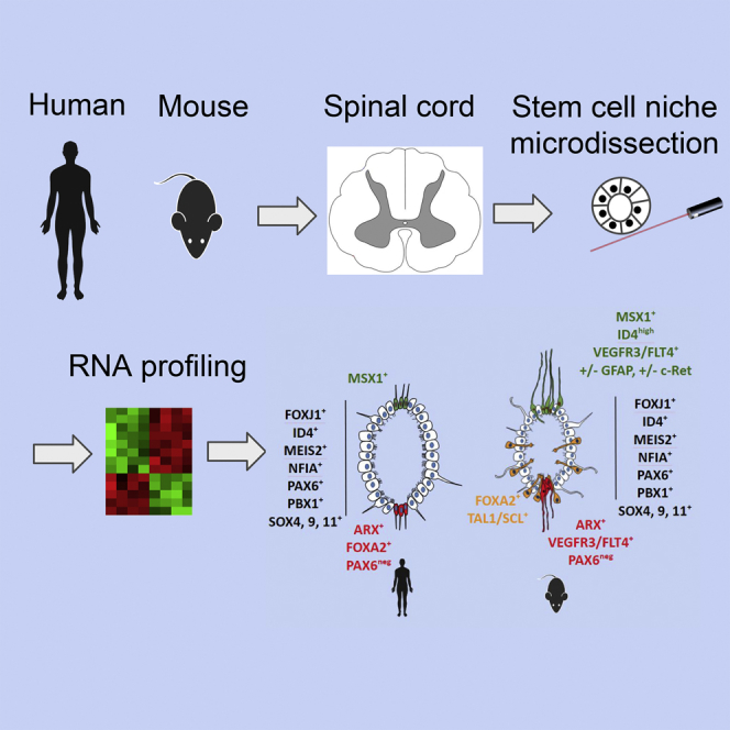
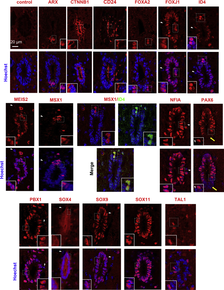
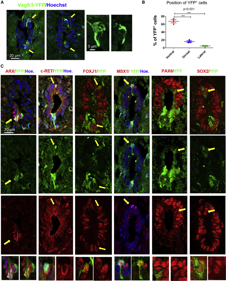
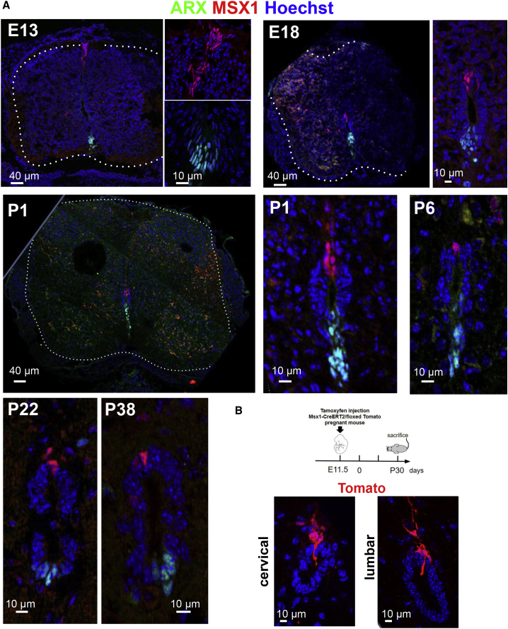
Anamniotes, rodents, and young humans maintain neural stem cells in the ependymal zone (EZ) around the central canal of the spinal cord, representing a possible endogenous source for repair in mammalian lesions. Cell diversity and genes specific for this region are ill defined. A cellular and molecular resource is provided here for the mouse and human EZ based on RNA profiling, immunostaining, and fluorescent transgenic mice. This uncovered the conserved expression of 1,200 genes including 120 transcription factors. Unexpectedly the EZ maintains an embryonic-like dorsal-ventral pattern of expression of spinal cord developmental transcription factors (ARX, FOXA2, MSX1, and PAX6). In mice, dorsal and ventral EZ cells express Vegfr3 and are derived from the embryonic roof and floor plates. The dorsal EZ expresses a high level of Bmp6 and Gdf10 genes and harbors a subpopulation of radial quiescent cells expressing MSX1 and ID4 transcription factors.

摘要

无脊椎动物、啮齿动物和幼年人类在脊髓中央管周围的室管膜区(EZ)中维持神经干细胞,这代表了哺乳动物病变中修复的可能内源性来源。该区域的细胞多样性和特定基因尚未明确界定。本文基于 RNA 分析、免疫染色和荧光转基因小鼠,为小鼠和人类 EZ 提供了一个细胞和分子资源。这揭示了包括 120 个转录因子在内的 1200 个基因的保守表达。出乎意料的是,EZ 维持了脊髓发育转录因子(ARX、FOXA2、MSX1 和 PAX6)的胚胎样背腹表达模式。在小鼠中,背侧和腹侧 EZ 细胞表达 Vegfr3,并且来源于胚胎的顶壁和基板。背侧 EZ 表达高水平的 Bmp6 和 Gdf10 基因,并含有一个表达 MSX1 和 ID4 转录因子的静止细胞亚群。

https://cdn.ncbi.nlm.nih.gov/pmc/blobs/31f4/6524006/ccd05b2baf78/gr6.jpg
https://cdn.ncbi.nlm.nih.gov/pmc/blobs/31f4/6524006/ac5e55667532/fx1.jpg
https://cdn.ncbi.nlm.nih.gov/pmc/blobs/31f4/6524006/7d29c3412057/gr1.jpg
https://cdn.ncbi.nlm.nih.gov/pmc/blobs/31f4/6524006/840febae28fb/gr2.jpg
https://cdn.ncbi.nlm.nih.gov/pmc/blobs/31f4/6524006/79a8acb1c9c7/gr3.jpg
https://cdn.ncbi.nlm.nih.gov/pmc/blobs/31f4/6524006/b70eb57cab7f/gr4.jpg
https://cdn.ncbi.nlm.nih.gov/pmc/blobs/31f4/6524006/29be482b8378/gr5.jpg
https://cdn.ncbi.nlm.nih.gov/pmc/blobs/31f4/6524006/ccd05b2baf78/gr6.jpg

相似文献

1
RNA Profiling of the Human and Mouse Spinal Cord Stem Cell Niches Reveals an Embryonic-like Regionalization with MSX1 Roof-Plate-Derived Cells.人类和小鼠脊髓干细胞龛的 RNA 分析揭示了具有 MSX1 顶盖板衍生细胞的胚胎样区域化。
Stem Cell Reports. 2019 May 14;12(5):1159-1177. doi: 10.1016/j.stemcr.2019.04.001. Epub 2019 Apr 25.
2
Persistence of FoxJ1 Pax6 Sox2 ependymal cells throughout life in the human spinal cord.FoxJ1、Pax6、Sox2 室管膜细胞在人类脊髓中的终生存在。
Cell Mol Life Sci. 2023 Jun 17;80(7):181. doi: 10.1007/s00018-023-04811-x.
3
A mesenchymal-like ZEB1(+) niche harbors dorsal radial glial fibrillary acidic protein-positive stem cells in the spinal cord.间质样 ZEB1(+)龛中存在背侧放射状胶质纤维酸性蛋白阳性的脊髓干细胞。
Stem Cells. 2009 Nov;27(11):2722-33. doi: 10.1002/stem.226.
4
Msx1 and Msx2 act as essential activators of Atoh1 expression in the murine spinal cord.Msx1 和 Msx2 作为 Atoh1 表达在小鼠脊髓中的必需激活物起作用。
Development. 2014 Apr;141(8):1726-36. doi: 10.1242/dev.099002.
5
Molecular mapping of the origin of postnatal spinal cord ependymal cells: evidence that adult ependymal cells are derived from Nkx6.1+ ventral neural progenitor cells.出生后脊髓室管膜细胞起源的分子图谱:成年室管膜细胞源自Nkx6.1+腹侧神经祖细胞的证据。
J Comp Neurol. 2003 Feb 10;456(3):237-44. doi: 10.1002/cne.10481.
6
Gliogenic radial glial cells show heterogeneity in the developing mouse spinal cord.神经胶质源性放射状胶质细胞在发育中的小鼠脊髓中表现出异质性。
Dev Neurosci. 2005;27(6):364-77. doi: 10.1159/000088452.
7
Cellular organization of the central canal ependymal zone, a niche of latent neural stem cells in the adult mammalian spinal cord.中枢管室管膜区的细胞组织,成年哺乳动物脊髓中潜在神经干细胞的龛位。
Neuroscience. 2009 Dec 15;164(3):1044-56. doi: 10.1016/j.neuroscience.2009.09.006. Epub 2009 Sep 9.
8
Multiple steps characterise ventricular layer attrition to form the ependymal cell lining of the adult mouse spinal cord central canal.心室层磨损形成成年小鼠脊髓中央管室管膜细胞 lining 需要经过多个步骤。
J Anat. 2020 Feb;236(2):334-350. doi: 10.1111/joa.13094. Epub 2019 Oct 31.
9
Floor plate descendants in the ependyma of the adult mouse Central Nervous System.成年小鼠中枢神经系统室管膜中底板衍生物
Int J Dev Biol. 2017;61(3-4-5):257-265. doi: 10.1387/ijdb.160232nb.
10
Spinal Cord Stem Cells In Their Microenvironment: The Ependyma as a Stem Cell Niche.脊髓干细胞及其微环境:室管膜作为干细胞龛。
Adv Exp Med Biol. 2017;1041:55-79. doi: 10.1007/978-3-319-69194-7_5.

引用本文的文献

1
Single-cell chromatin accessibility profiling of cell-state-specific gene regulatory programs during mouse organogenesis.小鼠器官发生过程中细胞状态特异性基因调控程序的单细胞染色质可及性分析
Front Neurosci. 2023 Jun 27;17:1170355. doi: 10.3389/fnins.2023.1170355. eCollection 2023.
2
The activation of dormant ependymal cells following spinal cord injury.脊髓损伤后静止的室管膜细胞的激活。
Stem Cell Res Ther. 2023 Jul 5;14(1):175. doi: 10.1186/s13287-023-03395-4.
3
Persistence of FoxJ1 Pax6 Sox2 ependymal cells throughout life in the human spinal cord.

本文引用的文献

1
Molecular Architecture of the Mouse Nervous System.小鼠神经系统的分子结构。
Cell. 2018 Aug 9;174(4):999-1014.e22. doi: 10.1016/j.cell.2018.06.021.
2
Wnt/β-catenin signaling regulates ependymal cell development and adult homeostasis.Wnt/β-catenin 信号通路调节室管膜细胞的发育和成年后的稳态。
Proc Natl Acad Sci U S A. 2018 Jun 26;115(26):E5954-E5962. doi: 10.1073/pnas.1803297115. Epub 2018 Jun 11.
3
The spinal ependymal zone as a source of endogenous repair cells across vertebrates.脊椎室管膜区作为脊椎动物内源性修复细胞的来源。
FoxJ1、Pax6、Sox2 室管膜细胞在人类脊髓中的终生存在。
Cell Mol Life Sci. 2023 Jun 17;80(7):181. doi: 10.1007/s00018-023-04811-x.
4
Endogenous neural stem cells characterization using omics approaches: Current knowledge in health and disease.使用组学方法表征内源性神经干细胞:健康与疾病的当前知识
Front Cell Neurosci. 2023 Apr 5;17:1125785. doi: 10.3389/fncel.2023.1125785. eCollection 2023.
5
Recent advances in understanding cell type transitions during dorsal neural tube development.背侧神经管发育过程中细胞类型转变认识的最新进展。
Fac Rev. 2022 Sep 27;11:27. doi: 10.12703/r/11-27. eCollection 2022.
6
DNGR-1-tracing marks an ependymal cell subset with damage-responsive neural stem cell potential.DNGR-1 示踪标记出具有损伤反应性神经干细胞潜能的室管膜细胞亚群。
Dev Cell. 2022 Aug 22;57(16):1957-1975.e9. doi: 10.1016/j.devcel.2022.07.012.
7
Stem Cell Therapies for Central Nervous System Trauma: The 4 Ws-What, When, Where, and Why.中枢神经系统创伤的干细胞治疗:4W-是什么、何时、何地和为什么。
Stem Cells Transl Med. 2022 Mar 3;11(1):14-25. doi: 10.1093/stcltm/szab006.
8
Regulating Endogenous Neural Stem Cell Activation to Promote Spinal Cord Injury Repair.调控内源性神经干细胞激活促进脊髓损伤修复。
Cells. 2022 Mar 1;11(5):846. doi: 10.3390/cells11050846.
9
RNA Profiling of Mouse Ependymal Cells after Spinal Cord Injury Identifies the Oncostatin Pathway as a Potential Key Regulator of Spinal Cord Stem Cell Fate.脊髓损伤后小鼠室管膜细胞的 RNA 谱分析鉴定出肿瘤坏死因子家族成员 11(oncostatin M,OSM)为脊髓干细胞命运的潜在关键调控因子。
Cells. 2021 Nov 27;10(12):3332. doi: 10.3390/cells10123332.
10
Positional Memory in Vertebrate Regeneration: A Century's Insights from the Salamander Limb.脊椎动物再生中的位置记忆:蝾螈肢体一个世纪的研究进展
Cold Spring Harb Perspect Biol. 2022 Jun 14;14(6):a040899. doi: 10.1101/cshperspect.a040899.
Prog Neurobiol. 2018 Nov;170:67-80. doi: 10.1016/j.pneurobio.2018.04.002. Epub 2018 Apr 9.
4
Single-cell profiling of the developing mouse brain and spinal cord with split-pool barcoding.单细胞分析发育中的鼠脑和脊髓的分裂池条形码技术。
Science. 2018 Apr 13;360(6385):176-182. doi: 10.1126/science.aam8999. Epub 2018 Mar 15.
5
Spinal Cord Stem Cells In Their Microenvironment: The Ependyma as a Stem Cell Niche.脊髓干细胞及其微环境:室管膜作为干细胞龛。
Adv Exp Med Biol. 2017;1041:55-79. doi: 10.1007/978-3-319-69194-7_5.
6
Ependymal cell contribution to scar formation after spinal cord injury is minimal, local and dependent on direct ependymal injury.室管膜细胞对脊髓损伤后瘢痕形成的贡献极小,局限于局部且依赖于直接的室管膜损伤。
Sci Rep. 2017 Jan 24;7:41122. doi: 10.1038/srep41122.
7
Msx1-Positive Progenitors in the Retinal Ciliary Margin Give Rise to Both Neural and Non-neural Progenies in Mammals.Msx1 阳性祖细胞在视网膜睫状缘产生哺乳动物的神经和非神经祖细胞。
Dev Cell. 2017 Jan 23;40(2):137-150. doi: 10.1016/j.devcel.2016.11.020. Epub 2016 Dec 20.
8
Adult NSC diversity and plasticity: the role of the niche.成体神经干细胞的多样性与可塑性:微环境的作用。
Curr Opin Neurobiol. 2017 Feb;42:68-74. doi: 10.1016/j.conb.2016.11.008. Epub 2016 Dec 12.
9
Myelin Basic Protein Regulates Primitive and Definitive Neural Stem Cell Proliferation from the Adult Spinal Cord.髓鞘碱性蛋白调节成体脊髓中原始和定型神经干细胞的增殖。
Stem Cells. 2017 Feb;35(2):485-496. doi: 10.1002/stem.2488. Epub 2016 Sep 23.
10
Floor plate descendants in the ependyma of the adult mouse Central Nervous System.成年小鼠中枢神经系统室管膜中底板衍生物
Int J Dev Biol. 2017;61(3-4-5):257-265. doi: 10.1387/ijdb.160232nb.