Women's Hospital, School of Medicine, Zhejiang University, Hangzhou, 310006, China.
J Ovarian Res. 2019 Apr 30;12(1):37. doi: 10.1186/s13048-019-0512-6.
The expression of PD-L1 has been reported in ovarian cancer. However, the prognostic role of PD-L1 expression in ovarian carcinoma remained controversial. This study was performed to assess the prognostic value of PD-L1 expression on ovarian cancer.
The PubMed, Embase, EBSCO, and Cochrane Library databases were searched to identify available publications. The pooled odds ratio (OR) or hazard ratios (HRs: multivariate analysis) with their 95% confidence intervals (95% CIs) were calculated in this analysis. A bioinformatics study based on The Cancer Genome Atlas (TCGA) sequencing and microarray datasets was used to further validate the results of PD-L1 mRNA expression. Kaplan-Meier (KM) survival curves were performed to evaluate the prognostic effect of PD-L1 mRNA expression.
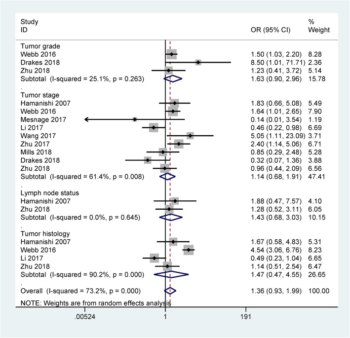
Twelve studies with 1630 ovarian cancers regarding PD-L1 immunohistochemical expression were identified. Meta-analysis showed that PD-L1 protein expression was not associated with tumor grade, clinical stage, lymph node status, tumor histology, overall survival (OS), and progression-free survival (PFS). TCGA data showed no association between PD-L1 mRNA expression and ovarian cancer. Further validation using microarray data suggested that no association between PD-L1 mRNA expression and OS was found in large independent patient cohorts (1310 cases). PD-L1 mRNA expression was significantly linked to worse PFS in 1228 patients with ovarian cancer (227458_at: HR = 1.55, 95% CI = 1.28-1.88, P < 0.001; 223834_at: HR = 1.41, 95% CI = 1.14-1.75, P = 0.0015).
Meta-analysis showed that PD-L1 may not be a prognostic factor for ovarian cancer. But a bioinformatics study showed that PD-L1 expression was significantly associated with worse PFS of ovarian cancer. More clinical studies are needed to further validate these findings.
PD-L1 的表达已在卵巢癌中报道。然而,PD-L1 表达在卵巢癌中的预后作用仍存在争议。本研究旨在评估 PD-L1 表达在卵巢癌中的预后价值。
检索 PubMed、Embase、EBSCO 和 Cochrane 图书馆数据库,以确定可用的出版物。在这项分析中,计算了合并的优势比 (OR) 或风险比 (HR:多变量分析) 及其 95%置信区间 (95%CI)。基于癌症基因组图谱 (TCGA) 测序和微阵列数据集的生物信息学研究用于进一步验证 PD-L1 mRNA 表达的结果。Kaplan-Meier (KM) 生存曲线用于评估 PD-L1 mRNA 表达的预后效应。
共有 12 项研究涉及 1630 例卵巢癌患者的 PD-L1 免疫组织化学表达,其中 12 项研究涉及 PD-L1 免疫组织化学表达。Meta 分析显示,PD-L1 蛋白表达与肿瘤分级、临床分期、淋巴结状态、肿瘤组织学、总生存 (OS) 和无进展生存 (PFS) 无关。TCGA 数据显示 PD-L1 mRNA 表达与卵巢癌无关。使用微阵列数据进行的进一步验证表明,在大型独立患者队列 (1310 例) 中,PD-L1 mRNA 表达与 OS 之间无关联。在 1228 例卵巢癌患者中,PD-L1 mRNA 表达与更差的 PFS 显著相关 (227458_at:HR=1.55,95%CI=1.28-1.88,P<0.001;223834_at:HR=1.41,95%CI=1.14-1.75,P=0.0015)。
Meta 分析表明,PD-L1 可能不是卵巢癌的预后因素。但一项生物信息学研究表明,PD-L1 表达与卵巢癌的更差 PFS 显著相关。需要更多的临床研究来进一步验证这些发现。