Bock Nathalie, Shokoohmand Ali, Kryza Thomas, Röhl Joan, Meijer Jonelle, Tran Phong A, Nelson Colleen C, Clements Judith A, Hutmacher Dietmar W
1School of Biomedical Sciences, Faculty of Health and Australian Prostate Cancer Research Centre (APCRC-Q), Institute of Health and Biomedical Innovation (IHBI), Queensland University of Technology (QUT), Brisbane, QLD 4000 Australia.
2Translational Research Institute (TRI), Woolloongabba, QLD 4102 Australia.
Bone Res. 2019 Apr 25;7:13. doi: 10.1038/s41413-019-0049-8. eCollection 2019.
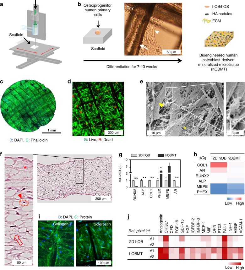
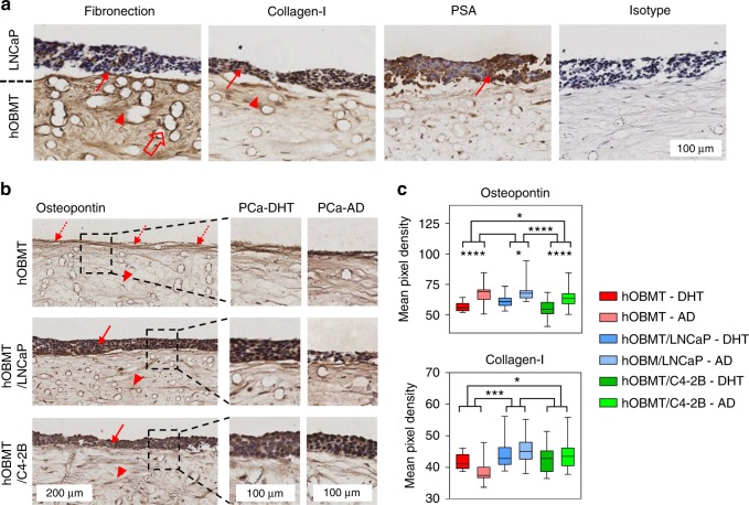
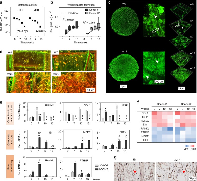
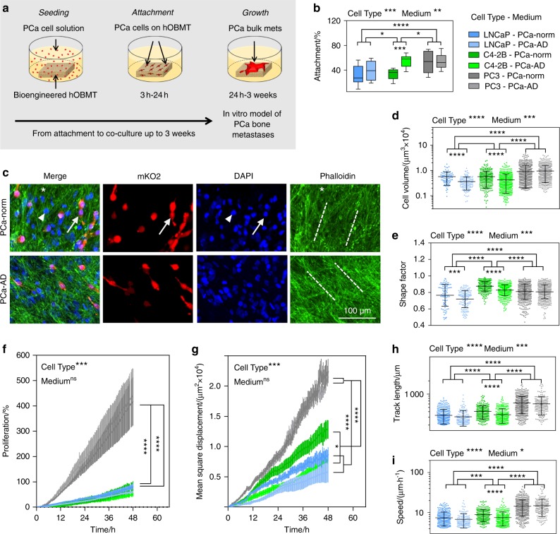
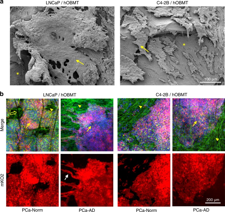
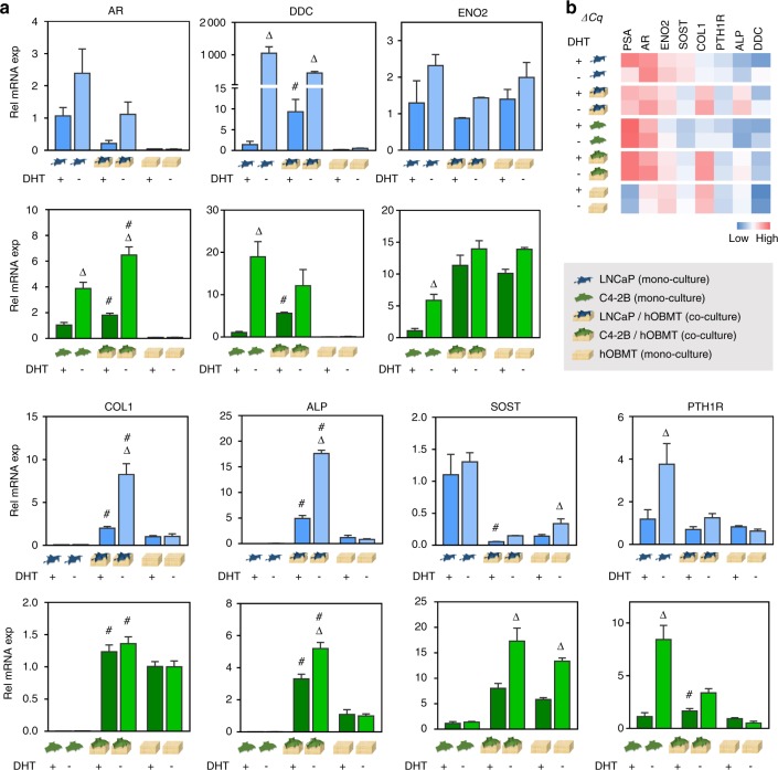
While stromal interactions are essential in cancer adaptation to hormonal therapies, the effects of bone stroma and androgen deprivation on cancer progression in bone are poorly understood. Here, we tissue-engineered and validated an in vitro microtissue model of osteoblastic bone metastases, and used it to study the effects of androgen deprivation in this microenvironment. The model was established by culturing primary human osteoprogenitor cells on melt electrowritten polymer scaffolds, leading to a mineralized osteoblast-derived microtissue containing, in a 3D setting, viable osteoblastic cells, osteocytic cells, and appropriate expression of osteoblast/osteocyte-derived mRNA and proteins, and mineral content. Direct co-culture of androgen receptor-dependent/independent cell lines (LNCaP, C4-2B, and PC3) led cancer cells to display functional and molecular features as observed in vivo. Co-cultured cancer cells showed increased affinity to the microtissues, as a function of their bone metastatic potential. Co-cultures led to alkaline phosphatase and collagen-I upregulation and sclerostin downregulation, consistent with the clinical marker profile of osteoblastic bone metastases. LNCaP showed a significant adaptive response under androgen deprivation in the microtissues, with the notable appearance of neuroendocrine transdifferentiation features and increased expression of related markers (dopa decarboxylase, enolase 2). Androgen deprivation affected the biology of the metastatic microenvironment with stronger upregulation of androgen receptor, alkaline phosphatase, and dopa decarboxylase, as seen in the transition towards resistance. The unique microtissues engineered here represent a substantial asset to determine the involvement of the human bone microenvironment in prostate cancer progression and response to a therapeutic context in this microenvironment.
虽然基质相互作用在癌症对激素疗法的适应性中至关重要,但骨基质和雄激素剥夺对骨中癌症进展的影响却知之甚少。在此,我们构建并验证了一种成骨细胞性骨转移的体外微组织模型,并利用它来研究雄激素剥夺在这种微环境中的作用。该模型通过将原代人骨祖细胞培养在熔喷电纺聚合物支架上建立,形成一种矿化的成骨细胞衍生微组织,在三维环境中包含有活力的成骨细胞、骨细胞,以及成骨细胞/骨细胞衍生的mRNA和蛋白质的适当表达和矿物质含量。雄激素受体依赖性/非依赖性细胞系(LNCaP、C4-2B和PC3)的直接共培养使癌细胞呈现出在体内观察到的功能和分子特征。共培养的癌细胞对微组织的亲和力增加,这是其骨转移潜能的一种表现。共培养导致碱性磷酸酶和I型胶原上调以及硬化蛋白下调,这与成骨细胞性骨转移的临床标志物特征一致。LNCaP在微组织中的雄激素剥夺条件下表现出显著的适应性反应,出现明显的神经内分泌转分化特征并增加相关标志物(多巴脱羧酶、烯醇化酶2)的表达。雄激素剥夺影响转移性微环境的生物学特性,雄激素受体、碱性磷酸酶和多巴脱羧酶的上调更强,这在向耐药性转变时可见。在此构建的独特微组织是确定人类骨微环境在前列腺癌进展中的作用以及对该微环境中治疗环境反应的重要资源。