• 文献检索
  • 文档翻译
  • 深度研究
  • 学术资讯
  • Suppr Zotero 插件Zotero 插件
  • 邀请有礼
  • 套餐&价格
  • 历史记录
应用&插件
Suppr Zotero 插件Zotero 插件浏览器插件Mac 客户端Windows 客户端微信小程序
定价
高级版会员购买积分包购买API积分包
服务
文献检索文档翻译深度研究API 文档MCP 服务
关于我们
关于 Suppr公司介绍联系我们用户协议隐私条款
关注我们

Suppr 超能文献

核心技术专利:CN118964589B侵权必究
粤ICP备2023148730 号-1Suppr @ 2026

文献检索

告别复杂PubMed语法,用中文像聊天一样搜索,搜遍4000万医学文献。AI智能推荐,让科研检索更轻松。

立即免费搜索

文件翻译

保留排版,准确专业,支持PDF/Word/PPT等文件格式,支持 12+语言互译。

免费翻译文档

深度研究

AI帮你快速写综述,25分钟生成高质量综述,智能提取关键信息,辅助科研写作。

立即免费体验

斜视性弱视患者脑区局部一致性改变的脑功能活动研究:静息态功能磁共振成像研究。

Altered brain activity in patients with strabismus and amblyopia detected by analysis of regional homogeneity: A resting‑state functional magnetic resonance imaging study.

机构信息

Department of Ophthalmology, The First Affiliated Hospital of Nanchang University, Jiangxi Province Ocular Disease Clinical Research Center, Nanchang, Jiangxi 330006, P.R. China.

Eye Institute of Xiamen University, Fujian Provincial Key Laboratory of Ophthalmology and Visual Science, Xiamen, Fujian 361000, P.R. China.

出版信息

Mol Med Rep. 2019 Jun;19(6):4832-4840. doi: 10.3892/mmr.2019.10147. Epub 2019 Apr 10.

DOI:10.3892/mmr.2019.10147
PMID:31059016
原文链接:https://pmc.ncbi.nlm.nih.gov/articles/PMC6522834/
Abstract

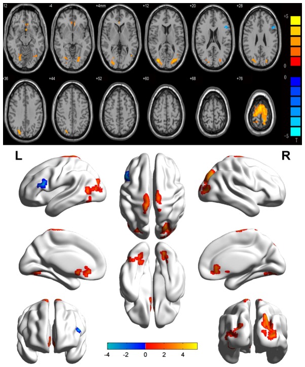
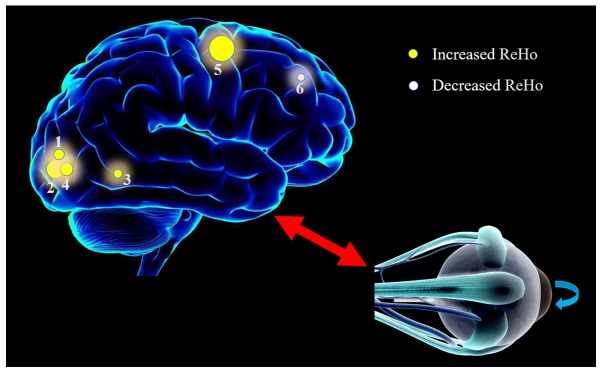
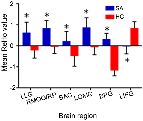
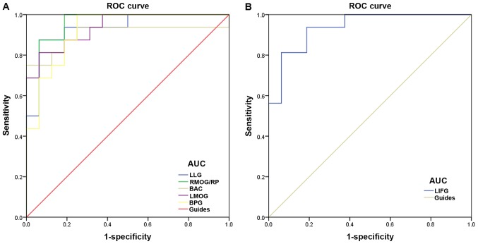
Previous studies have demonstrated that strabismus or amblyopia can result in marked brain function and anatomical alterations. However, differences in spontaneous brain activity in strabismus and amblyopia (SA) patients as compared with control individuals remain unclear. The present study aimed to analyze the potential brain activity changes in SA patients and their association with behavioral performance. In total, 16 patients with SA (10 women and 6 men) and 16 healthy controls (HCs; 6 men and 10 women) with matched age and sex were recruited. All subjects were examined with resting‑state functional magnetic resonance imaging (rs‑fMRI), and changes in the spontaneous brain activity of SA patients were evaluated by the regional homogeneity (ReHo) method. The diagnostic ability of the ReHo method was assessed using receiver operating characteristic (ROC) curve analysis. In addition, the association between the mean ReHo value in different brain regions and the behavioral performance was explored by correlation analysis. It was observed that the ReHo value was significantly increased in SA patients compared with HCs in the following brain regions: left lingual gyrus, right middle occipital gyrus/precuneus, bilateral anterior cingulate, left middle occipital gyrus and bilateral precentral gyrus. By contrast, the ReHo value of the left inferior frontal gyrus was significantly lower than that in HCs. ROC curve analysis indicated that the ReHo method has certain credibility for the diagnosis of SA patients. In addition, no similar changes were detected in other brain regions. These results revealed abnormal spontaneous brain activity in certain parts of the brain in adult patients with SA, which suggests the involvement of the neuropathological or compensatory mechanism in these patients, and may be beneficial for clinical treatment.

摘要

先前的研究表明,斜视或弱视会导致明显的大脑功能和解剖结构改变。然而,斜视和弱视(SA)患者与正常对照个体之间自发脑活动的差异尚不清楚。本研究旨在分析 SA 患者潜在的脑活动变化及其与行为表现的关系。共纳入 16 例 SA 患者(10 名女性和 6 名男性)和 16 名健康对照者(HCs;6 名男性和 10 名女性),两组年龄和性别相匹配。所有受试者均接受静息态功能磁共振成像(rs-fMRI)检查,采用局部一致性(ReHo)方法评估 SA 患者自发脑活动的变化。采用受试者工作特征(ROC)曲线分析评估 ReHo 方法的诊断能力。此外,通过相关性分析探讨了不同脑区平均 ReHo 值与行为表现之间的关系。结果显示,与 HCs 相比,SA 患者的 ReHo 值在以下脑区显著升高:左侧舌回、右侧中枕叶/楔前叶、双侧前扣带回、左侧中枕叶和双侧中央前回。相比之下,左侧额下回的 ReHo 值明显低于 HCs。ROC 曲线分析表明,ReHo 方法对 SA 患者的诊断具有一定的可信度。此外,在其他脑区未发现类似的变化。这些结果表明,SA 成年患者某些脑区存在异常自发脑活动,提示这些患者存在神经病理学或代偿机制参与,可能有助于临床治疗。

https://cdn.ncbi.nlm.nih.gov/pmc/blobs/885b/6522834/92a7cb5b8999/MMR-19-06-4832-g03.jpg
https://cdn.ncbi.nlm.nih.gov/pmc/blobs/885b/6522834/9d65981f8cee/MMR-19-06-4832-g00.jpg
https://cdn.ncbi.nlm.nih.gov/pmc/blobs/885b/6522834/eb38714e1634/MMR-19-06-4832-g01.jpg
https://cdn.ncbi.nlm.nih.gov/pmc/blobs/885b/6522834/771b44f12acb/MMR-19-06-4832-g02.jpg
https://cdn.ncbi.nlm.nih.gov/pmc/blobs/885b/6522834/92a7cb5b8999/MMR-19-06-4832-g03.jpg

相似文献

1
Altered brain activity in patients with strabismus and amblyopia detected by analysis of regional homogeneity: A resting‑state functional magnetic resonance imaging study.斜视性弱视患者脑区局部一致性改变的脑功能活动研究:静息态功能磁共振成像研究。
Mol Med Rep. 2019 Jun;19(6):4832-4840. doi: 10.3892/mmr.2019.10147. Epub 2019 Apr 10.
2
Altered Brain Network Centrality in Patients with Adult Strabismus with Amblyopia: A Resting-State Functional Magnetic Resonance Imaging (fMRI) Study.成人斜视性弱视患者脑网络中心性改变:一项静息态功能磁共振成像(fMRI)研究。
Med Sci Monit. 2020 Nov 23;26:e925856. doi: 10.12659/MSM.925856.
3
Altered Brain Activity in Strabismic Amblyopic Children as Determined by Regional Homogeneity: A Resting-State Functional Magnetic Resonance Imaging Study.基于局部一致性测定的斜视性弱视儿童脑活动改变:一项静息态功能磁共振成像研究
Front Neurosci. 2022 Jun 2;16:879253. doi: 10.3389/fnins.2022.879253. eCollection 2022.
4
Altered regional homogeneity in patients with late monocular blindness: a resting-state functional MRI study.晚期单眼失明患者的局部一致性改变:一项静息态功能磁共振成像研究
Neuroreport. 2017 Nov 8;28(16):1085-1091. doi: 10.1097/WNR.0000000000000855.
5
Altered regional homogeneity of brain spontaneous signals in SIV infected rhesus macaque model.感染猴免疫缺陷病毒的恒河猴模型中脑自发信号的局部一致性改变
Magn Reson Imaging. 2017 Apr;37:56-61. doi: 10.1016/j.mri.2016.10.019. Epub 2016 Oct 27.
6
Altered Spontaneous Brain Activity in Patients with Classical Trigeminal Neuralgia Using Regional Homogeneity: A Resting-State Functional MRI Study.基于局部一致性分析的原发性三叉神经痛患者自发性脑活动改变:一项静息态功能磁共振研究。
Pain Pract. 2019 Apr;19(4):397-406. doi: 10.1111/papr.12753. Epub 2019 Jan 25.
7
Abnormalities of regional homogeneity and its correlation with clinical symptoms in Naïve patients with first-episode schizophrenia.首发精神分裂症患者静息态局部一致性改变及其与临床症状的相关性
Brain Imaging Behav. 2019 Apr;13(2):503-513. doi: 10.1007/s11682-018-9882-4.
8
Altered spontaneous brain activity in patients with strabismic amblyopia: A resting-state fMRI study using regional homogeneity analysis.斜视性弱视患者自发脑活动的改变:一项使用局部一致性分析的静息态功能磁共振成像研究
Exp Ther Med. 2019 Nov;18(5):3877-3884. doi: 10.3892/etm.2019.8038. Epub 2019 Sep 23.
9
Gray matter volume alterations in patients with strabismus and amblyopia: voxel-based morphometry study.斜视性弱视患者脑灰质体积改变的基于体素的形态测量学研究。
Sci Rep. 2022 Jan 10;12(1):458. doi: 10.1038/s41598-021-04184-w.
10
ALTERED BRAIN ACTIVITY IN PATIENTS WITH DIABETIC RETINOPATHY USING REGIONAL HOMOGENEITY: A RESTING-STATE fMRI STUDY.糖尿病视网膜病变患者局部一致性的脑活动改变:一项静息态 fMRI 研究。
Endocr Pract. 2019 Apr;25(4):320-327. doi: 10.4158/EP-2018-0517.

引用本文的文献

1
Machine Learning-Based Identification of Children With Intermittent Exotropia Using Multiple Resting-State Functional Magnetic Resonance Imaging Features.基于机器学习利用多个静息态功能磁共振成像特征识别间歇性外斜视儿童
Brain Behav. 2025 May;15(5):e70556. doi: 10.1002/brb3.70556.
2
Brain activity in different brain areas of patients with diabetic vitreous hemorrhage according to voxel-based morphometry.基于体素形态测量法的糖尿病性玻璃体出血患者不同脑区的脑活动情况
Int J Ophthalmol. 2025 Feb 18;18(2):258-267. doi: 10.18240/ijo.2025.02.09. eCollection 2025.
3
Altered spontaneous brain activity in children with deprivation amblyopia: a resting-state functional magnetic resonance imaging study.

本文引用的文献

1
Abnormal regional spontaneous neural activity in visual pathway in retinal detachment patients: a resting-state functional MRI study.视网膜脱离患者视觉通路区域自发性神经活动异常:一项静息态功能磁共振成像研究。
Neuropsychiatr Dis Treat. 2017 Nov 22;13:2849-2854. doi: 10.2147/NDT.S147645. eCollection 2017.
2
Duane retraction syndrome: causes, effects and management strategies.杜安眼球后退综合征:病因、影响及治疗策略。
Clin Ophthalmol. 2017 Oct 30;11:1917-1930. doi: 10.2147/OPTH.S127481. eCollection 2017.
3
Altered regional homogeneity in patients with late monocular blindness: a resting-state functional MRI study.
剥夺性弱视儿童的自发脑活动改变:一项静息态功能磁共振成像研究
Eur J Med Res. 2025 Jan 15;30(1):31. doi: 10.1186/s40001-025-02275-2.
4
Abnormal topological organization of functional brain networks in the patients with anterior segment ischemic optic neuropathy.前段缺血性视神经病变患者功能性脑网络的异常拓扑组织
Front Neurosci. 2024 Nov 22;18:1458897. doi: 10.3389/fnins.2024.1458897. eCollection 2024.
5
The Impact of the Wavelength and Its Transmittance on the Visual Evoked Potentials, at Baseline, and under the Effect of Six Monochromatic Filters Used for Visual Treatments.波长及其透过率对视觉诱发电位的影响,在基线时和在用于视觉治疗的六个单色滤光片的作用下。
Sensors (Basel). 2023 May 31;23(11):5227. doi: 10.3390/s23115227.
6
Structural and functional alterations in the brains of patients with anisometropic and strabismic amblyopia: a systematic review of magnetic resonance imaging studies.屈光参差性和斜视性弱视患者大脑的结构和功能改变:磁共振成像研究的系统评价
Neural Regen Res. 2023 Nov;18(11):2348-2356. doi: 10.4103/1673-5374.371349.
7
Continuous and intermittent theta burst stimulation of primary visual cortex do not modulate resting state functional connectivity: A sham-controlled multi-echo fMRI study.持续和间断 theta 爆发刺激初级视觉皮层不会调节静息状态功能连接:一项 sham 对照多回波 fMRI 研究。
Brain Behav. 2023 May;13(5):e2989. doi: 10.1002/brb3.2989. Epub 2023 Apr 16.
8
Comitant strabismus etiology: extraocular muscle integrity and central nervous system involvement-a narrative review.共同性斜视病因:眼外肌完整性和中枢神经系统受累——叙述性综述。
Graefes Arch Clin Exp Ophthalmol. 2023 Jul;261(7):1781-1792. doi: 10.1007/s00417-022-05935-9. Epub 2023 Jan 21.
9
Differences in the Visual Performances of Patients with Strabismus, Amblyopia, and Healthy Controls.斜视、弱视患者与健康对照者视觉表现的差异。
Bioengineering (Basel). 2022 Oct 29;9(11):626. doi: 10.3390/bioengineering9110626.
10
Amblyopia: progress and promise of functional magnetic resonance imaging.弱视:功能磁共振成像的进展与前景
Graefes Arch Clin Exp Ophthalmol. 2023 May;261(5):1229-1246. doi: 10.1007/s00417-022-05826-z. Epub 2022 Oct 25.
晚期单眼失明患者的局部一致性改变:一项静息态功能磁共振成像研究
Neuroreport. 2017 Nov 8;28(16):1085-1091. doi: 10.1097/WNR.0000000000000855.
4
The atrophy of white and gray matter volume in patients with comitant strabismus: Evidence from a voxel-based morphometry study.伴有斜视患者白质和灰质体积萎缩:基于体素的形态学研究证据。
Mol Med Rep. 2017 Sep;16(3):3276-3282. doi: 10.3892/mmr.2017.7006. Epub 2017 Jul 15.
5
Impaired Activation of Visual Attention Network for Motion Salience Is Accompanied by Reduced Functional Connectivity between Frontal Eye Fields and Visual Cortex in Strabismic Amblyopia.斜视性弱视中,视觉注意网络对运动显著性的激活受损,同时额眼区与视觉皮层之间的功能连接减少。
Front Hum Neurosci. 2017 Apr 21;11:195. doi: 10.3389/fnhum.2017.00195. eCollection 2017.
6
Altered interhemispheric functional connectivity in patients with anisometropic and strabismic amblyopia: a resting-state fMRI study.屈光参差性和斜视性弱视患者半球间功能连接的改变:一项静息态功能磁共振成像研究
Neuroradiology. 2017 May;59(5):517-524. doi: 10.1007/s00234-017-1824-0. Epub 2017 Mar 24.
7
Microstructural changes of the whole brain in patients with comitant strabismus: evidence from a diffusion tensor imaging study.共同性斜视患者全脑的微观结构变化:来自扩散张量成像研究的证据
Neuropsychiatr Dis Treat. 2016 Aug 12;12:2007-14. doi: 10.2147/NDT.S108834. eCollection 2016.
8
Altered regional homogeneity in patients with unilateral acute open-globe injury: a resting-state functional MRI study.单侧急性开放性眼球损伤患者的局部一致性改变:一项静息态功能磁共振成像研究。
Neuropsychiatr Dis Treat. 2016 Aug 1;12:1901-6. doi: 10.2147/NDT.S110541. eCollection 2016.
9
Altered intrinsic regional brain spontaneous activity in patients with comitant strabismus: a resting-state functional MRI study.共同性斜视患者脑内固有区域自发活动的改变:一项静息态功能磁共振成像研究
Neuropsychiatr Dis Treat. 2016 Jun 3;12:1303-8. doi: 10.2147/NDT.S105478. eCollection 2016.
10
Association of Optic Radiation Integrity with Cortical Thickness in Children with Anisometropic Amblyopia.屈光不正性弱视儿童视辐射完整性与皮质厚度的相关性研究。
Neurosci Bull. 2016 Feb;32(1):51-60. doi: 10.1007/s12264-015-0005-6. Epub 2016 Jan 14.