Reich Michael, Cakir Bertan, Cvetkoski Stevan, Lang Stefan J, Stahl Andreas, Ness Thomas, Agostini Hansjürgen, Lange Clemens
Eye Center, Faculty of Medicine, Albert-Ludwigs University Freiburg, Killianstrasse 5, 79106, Freiburg, Germany.
Clinic for General and Visceral Surgery, Loerrach, Germany.
BMC Ophthalmol. 2019 May 7;19(1):104. doi: 10.1186/s12886-019-1111-4.
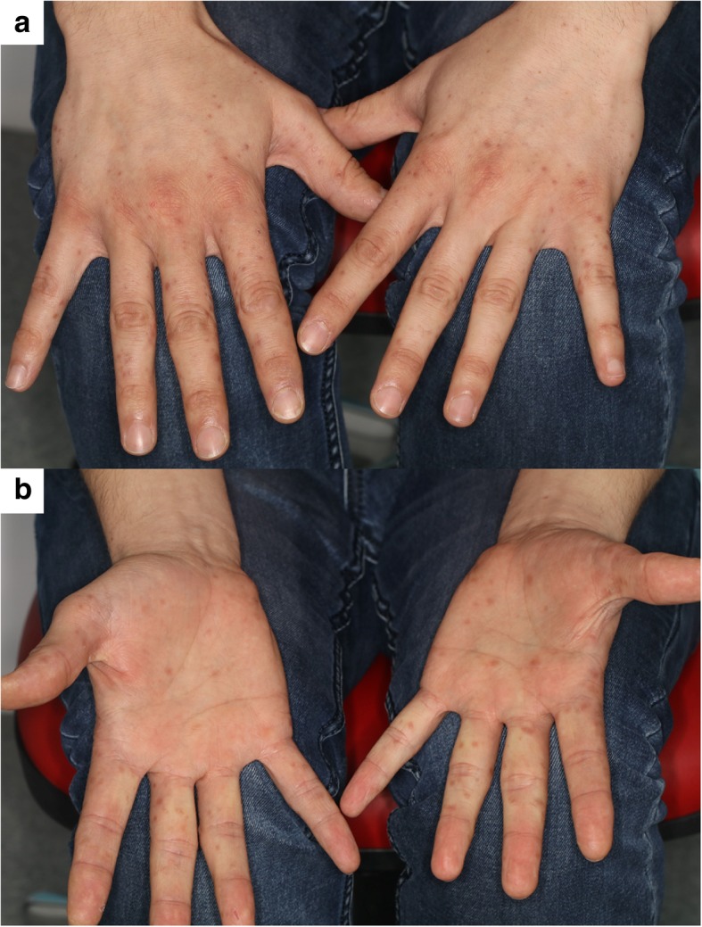
To report the case of a 31-year-old patient with Hand, Foot and Mouth Disease (HFMD) and concurrent acute monocular maculopathy, and to describe multimodal imaging findings never before described including optical coherence tomography angiography (OCT-A).
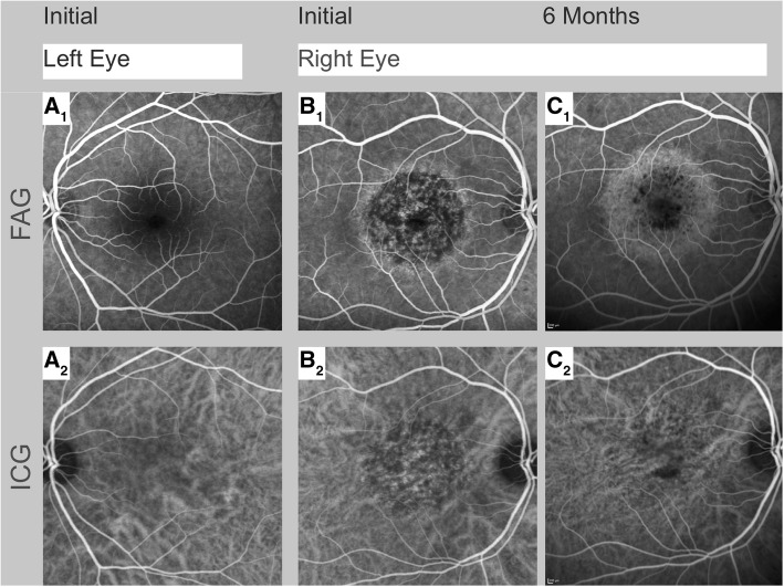
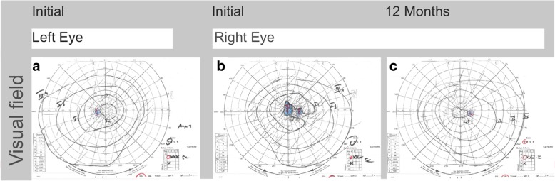
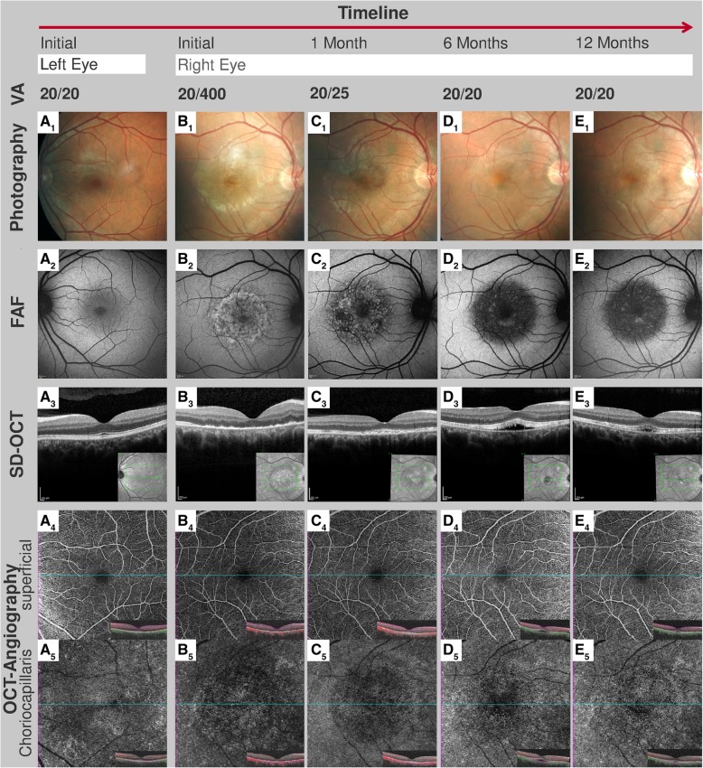
Nine days after the onset of clinically highly probable but not laboratory-verified HFMD, a 31-year old male noticed a central scotoma, distorted lines and loss of visual acuity (Snellen visual acuity 20/400) in his right eye. Funduscopy revealed focal alterations in the retinal pigmented epithelium (RPE) and yellow retinal dots corresponding to focal dots of decreased fundus autofluorescence (FAF) surrounded by increased FAF. Spectral domain optical coherence tomography (SD-OCT) demonstrated irregularities in the ellipsoide zone, hyperreflective dots above the RPE and RPE thickening. Fundus fluorescein angiography (FAG) revealed central hypofluorescence in the macular area in the early phase, as well as increasing focal hyperfluorescence in the late phase corresponding with RPE defects observed in FAF. Indocyanine green angiography (ICGA) showed central hypofluorescence in the early and late phase, corresponding with areas of reduced flow in the choroidea and choriocapillaris as apparent in OCT-A. Visual acuity improved within 3 months without any systemic or local therapy. At his three-month follow-up, SD-OCT revealed subtle subretinal fluid that resolved spontaneously over time. No signs of choroidal neovascularization were observed. Twelve months following the onset of symptoms Snellen visual acuity was 400/400. Multimodal imaging revealed subtly changed, decreased FAF while the choroidal architecture recovered completely as demonstrated by OCT-A.
HFMD-associated maculopahty is an uncommon but important differential diagnosis of chorioretinitis with macular involvement. The prognosis can be good and the initially observed morphological pathologies such as impaired perfusion of the choroidal vessels can recover spontaneously over a period lasting 12 months. OCT-A can be employed as a non-invasive tool to detect the reduced perfusion of the choroidal vessels and for monitoring the disease course.
报告一名31岁手足口病(HFMD)患者并发急性单眼黄斑病变的病例,并描述包括光学相干断层扫描血管造影(OCT-A)在内的此前从未描述过的多模态成像结果。
在临床高度疑似但未经实验室确诊的手足口病发病九天后,一名31岁男性注意到其右眼出现中心暗点、线条扭曲及视力下降(Snellen视力20/400)。眼底镜检查发现视网膜色素上皮(RPE)有局灶性改变,以及与眼底自发荧光(FAF)降低的局灶性小点相对应的黄色视网膜小点,周围FAF增强。频域光学相干断层扫描(SD-OCT)显示椭圆体带不规则、RPE上方的高反射小点及RPE增厚。眼底荧光血管造影(FAG)显示早期黄斑区中心低荧光,晚期局灶性高荧光增加,与FAF中观察到的RPE缺损相对应。吲哚菁绿血管造影(ICGA)显示早期和晚期中心低荧光,与OCT-A中脉络膜和脉络膜毛细血管血流减少的区域相对应。未经任何全身或局部治疗,视力在3个月内有所改善。在三个月随访时,SD-OCT显示有细微的视网膜下液,随时间自发消退。未观察到脉络膜新生血管的迹象。症状出现十二个月后,Snellen视力为400/400。多模态成像显示FAF略有变化且降低,而脉络膜结构如OCT-A所示已完全恢复。
手足口病相关黄斑病变是黄斑受累的脉络膜视网膜炎一种罕见但重要的鉴别诊断。预后可能良好,最初观察到的形态学病变如脉络膜血管灌注受损可在12个月内自发恢复。OCT-A可作为一种非侵入性工具,用于检测脉络膜血管灌注减少及监测疾病进程。