MedStar Georgetown University Hospital, 3800 Reservoir Road, NW CG201, Washington, DC, 20007, USA.
J Digit Imaging. 2019 Aug;32(4):656-664. doi: 10.1007/s10278-019-00226-y.
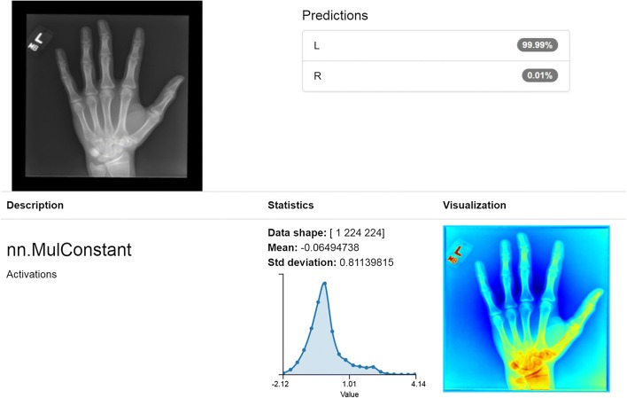
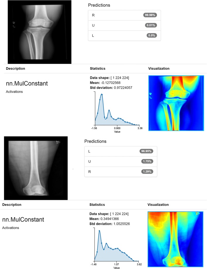
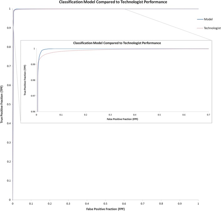
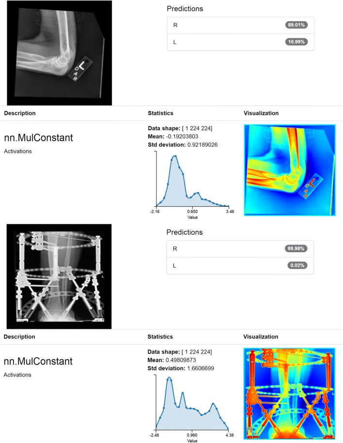
Develop a highly accurate deep learning model to reliably classify radiographs by laterality. Digital Imaging and Communications in Medicine (DICOM) data for nine body parts was extracted retrospectively. Laterality was determined directly if encoded properly or inferred using other elements. Curation confirmed categorization and identified inaccurate labels due to human error. Augmentation enriched training data to semi-equilibrate classes. Classification and object detection models were developed on a dedicated workstation and tested on novel images. Receiver operating characteristic (ROC) curves, sensitivity, specificity, and accuracy were calculated. Study-level accuracy was determined and both were compared to human performance. An ensemble model was tested for the rigorous use-case of automatically classifying exams retrospectively. The final classification model identified novel images with an ROC area under the curve (AUC) of 0.999, improving on previous work and comparable to human performance. A similar ROC curve was observed for per-study analysis with AUC of 0.999. The object detection model classified images with accuracy of 99% or greater at both image and study level. Confidence scores allow adjustment of sensitivity and specificity as needed; the ensemble model designed for the highly specific use-case of automatically classifying exams was comparable and arguably better than human performance demonstrating 99% accuracy with 1% of exams unchanged and no incorrect classification. Deep learning models can classify radiographs by laterality with high accuracy and may be applied in a variety of settings that could improve patient safety and radiologist satisfaction. Rigorous use-cases requiring high specificity are achievable.
开发一种高度准确的深度学习模型,以可靠地对放射线照片进行侧别分类。回顾性提取了九个身体部位的数字成像和通信医学(DICOM)数据。如果编码正确,则直接确定侧别,否则使用其他元素推断侧别。策展确认了分类,并确定了由于人为错误导致的不准确标签。扩充丰富了训练数据,以实现半均衡化分类。在专用工作站上开发分类和目标检测模型,并在新图像上进行测试。计算了接收者操作特征(ROC)曲线、敏感性、特异性和准确性。确定了研究级别的准确性,并将其与人类性能进行了比较。还针对自动回顾性分类考试的严格用例测试了集成模型。最终的分类模型对新图像的 ROC 曲线下面积(AUC)为 0.999,优于先前的工作,且与人类性能相当。对每项研究的分析也观察到类似的 ROC 曲线,AUC 为 0.999。目标检测模型在图像和研究级别上的准确率均达到 99%或更高。置信度分数允许根据需要调整敏感性和特异性;为高度特定的用例(即自动分类考试)设计的集成模型与人类性能相当,甚至可能更好,证明准确率为 99%,有 1%的考试不变且无错误分类。深度学习模型可以高精度地对放射线照片进行侧别分类,并且可以应用于各种可能提高患者安全和放射科医生满意度的环境中。可以实现需要高特异性的严格用例。