• 文献检索
  • 文档翻译
  • 深度研究
  • 学术资讯
  • Suppr Zotero 插件Zotero 插件
  • 邀请有礼
  • 套餐&价格
  • 历史记录
应用&插件
Suppr Zotero 插件Zotero 插件浏览器插件Mac 客户端Windows 客户端微信小程序
定价
高级版会员购买积分包购买API积分包
服务
文献检索文档翻译深度研究API 文档MCP 服务
关于我们
关于 Suppr公司介绍联系我们用户协议隐私条款
关注我们

Suppr 超能文献

核心技术专利:CN118964589B侵权必究
粤ICP备2023148730 号-1Suppr @ 2026

文献检索

告别复杂PubMed语法,用中文像聊天一样搜索,搜遍4000万医学文献。AI智能推荐,让科研检索更轻松。

立即免费搜索

文件翻译

保留排版,准确专业,支持PDF/Word/PPT等文件格式,支持 12+语言互译。

免费翻译文档

深度研究

AI帮你快速写综述,25分钟生成高质量综述,智能提取关键信息,辅助科研写作。

立即免费体验

不对称募集和肌动蛋白依赖性皮层流驱动神经母细胞极性循环。

Asymmetric recruitment and actin-dependent cortical flows drive the neuroblast polarity cycle.

机构信息

Department of Chemistry and Biochemistry, Institute of Molecular Biology, University of Oregon, Eugene, United States.

出版信息

Elife. 2019 May 8;8:e45815. doi: 10.7554/eLife.45815.

DOI:10.7554/eLife.45815
PMID:31066675
原文链接:https://pmc.ncbi.nlm.nih.gov/articles/PMC6524966/
Abstract

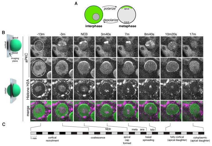
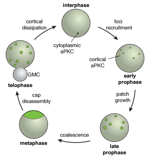
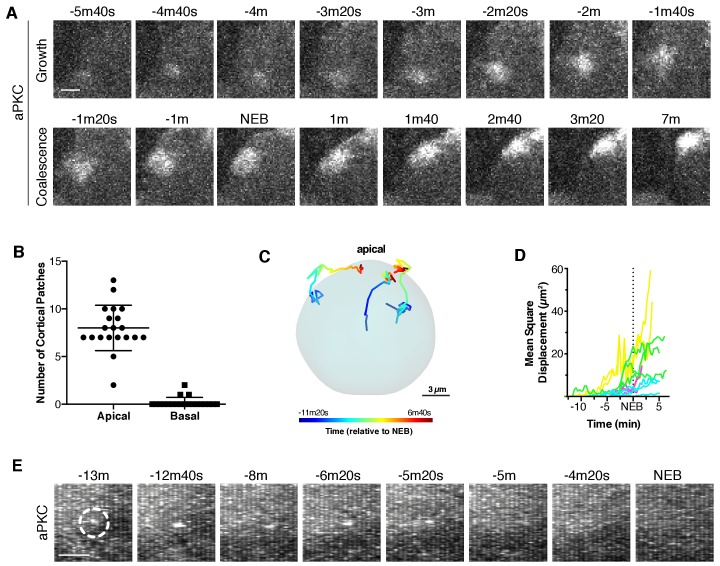
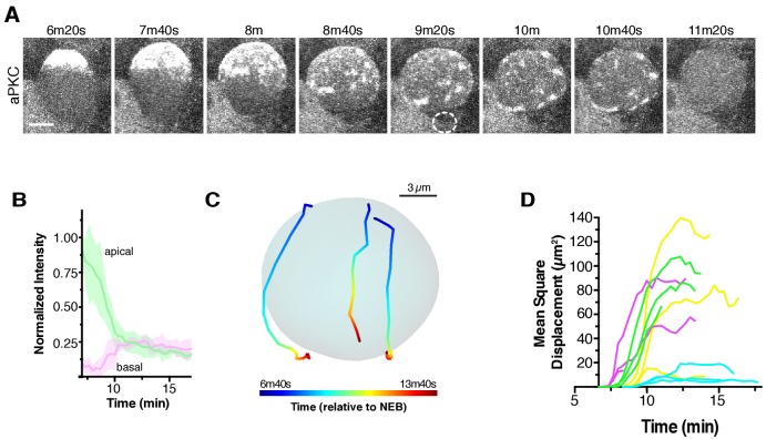
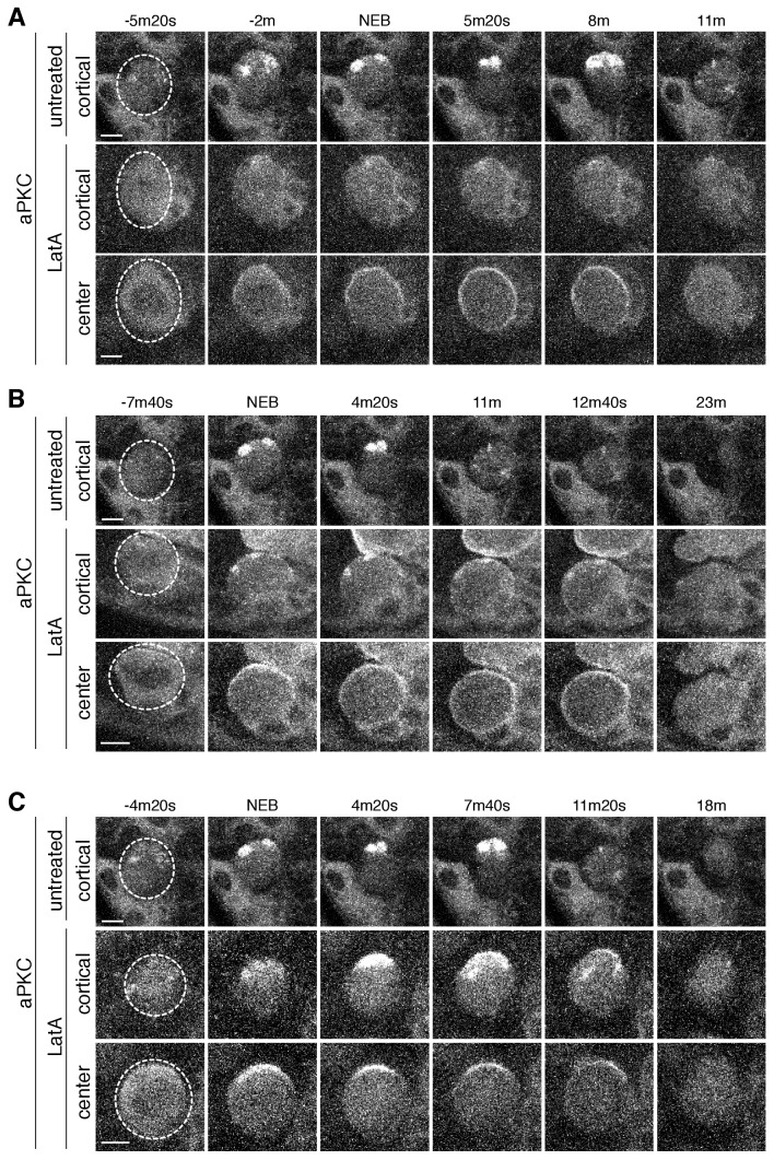
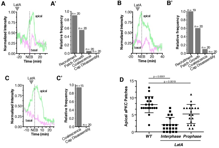
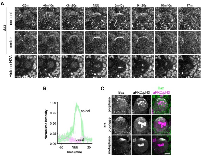
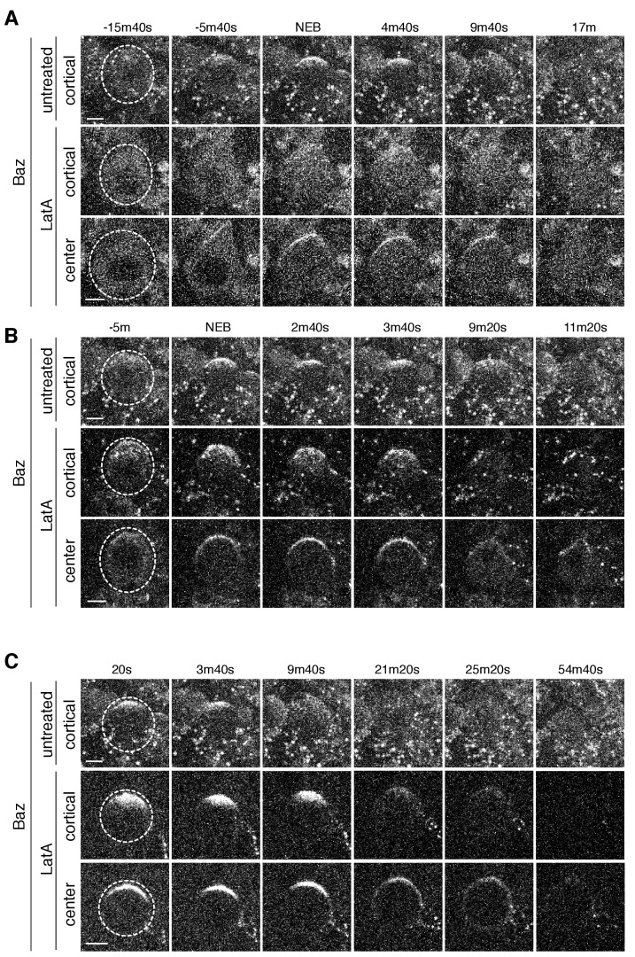
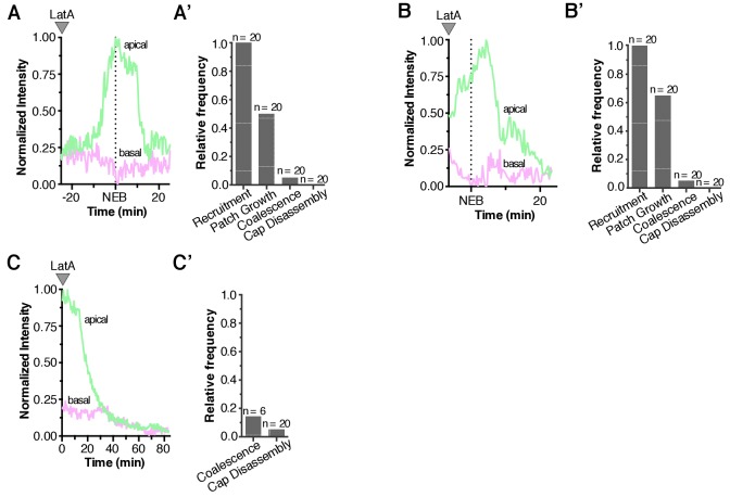
During the asymmetric divisions of neuroblasts, the Par polarity complex cycles between the cytoplasm and an apical cortical domain that restricts differentiation factors to the basal cortex. We used rapid imaging of the full cell volume to uncover the dynamic steps that underlie transitions between neuroblast polarity states. Initially, the Par proteins aPKC and Bazooka form discrete foci at the apical cortex. Foci grow into patches that together comprise a discontinuous, unorganized structure. Coordinated cortical flows that begin near metaphase and are dependent on the actin cytoskeleton rapidly transform the patches into a highly organized apical cap. At anaphase onset, the cap disassembles as the cortical flow reverses direction toward the emerging cleavage furrow. Following division, cortical patches dissipate into the cytoplasm allowing the neuroblast polarity cycle to begin again. Our work demonstrates how neuroblasts use asymmetric recruitment and cortical flows to dynamically polarize during asymmetric division cycles.

摘要

在神经母细胞的不对称分裂过程中,Par 极性复合物在细胞质和限制分化因子到基底皮质的顶端皮质域之间循环。我们使用全细胞体积的快速成像来揭示神经母细胞极性状态转变背后的动态步骤。最初,Par 蛋白 aPKC 和 Bazooka 在顶端皮质形成离散焦点。焦点生长成斑块,共同组成一个不连续的、无组织的结构。从中期开始并依赖肌动蛋白细胞骨架的协调皮层流迅速将斑块转化为高度组织化的顶端帽。在有丝分裂开始时,帽随着皮层流向新出现的分裂沟的方向反转而解体。分裂后,皮质斑块消散到细胞质中,允许神经母细胞极性循环再次开始。我们的工作展示了神经母细胞如何在不对称分裂周期中利用不对称募集和皮层流来动态极化。

https://cdn.ncbi.nlm.nih.gov/pmc/blobs/46b2/6524966/d5ad14ab2bcc/elife-45815-fig7.jpg
https://cdn.ncbi.nlm.nih.gov/pmc/blobs/46b2/6524966/2217173f7506/elife-45815-fig1.jpg
https://cdn.ncbi.nlm.nih.gov/pmc/blobs/46b2/6524966/788d903d579e/elife-45815-fig1-figsupp1.jpg
https://cdn.ncbi.nlm.nih.gov/pmc/blobs/46b2/6524966/602706e1ee91/elife-45815-fig2.jpg
https://cdn.ncbi.nlm.nih.gov/pmc/blobs/46b2/6524966/5ec77f2fdfbe/elife-45815-fig3.jpg
https://cdn.ncbi.nlm.nih.gov/pmc/blobs/46b2/6524966/fdc79db450f1/elife-45815-fig4.jpg
https://cdn.ncbi.nlm.nih.gov/pmc/blobs/46b2/6524966/bc49dc8a5c33/elife-45815-fig4-figsupp1.jpg
https://cdn.ncbi.nlm.nih.gov/pmc/blobs/46b2/6524966/1e4f105dc667/elife-45815-fig5.jpg
https://cdn.ncbi.nlm.nih.gov/pmc/blobs/46b2/6524966/7fffaa137947/elife-45815-fig6.jpg
https://cdn.ncbi.nlm.nih.gov/pmc/blobs/46b2/6524966/97f59705def1/elife-45815-fig6-figsupp1.jpg
https://cdn.ncbi.nlm.nih.gov/pmc/blobs/46b2/6524966/d5ad14ab2bcc/elife-45815-fig7.jpg

相似文献

1
Asymmetric recruitment and actin-dependent cortical flows drive the neuroblast polarity cycle.不对称募集和肌动蛋白依赖性皮层流驱动神经母细胞极性循环。
Elife. 2019 May 8;8:e45815. doi: 10.7554/eLife.45815.
2
Phases of cortical actomyosin dynamics coupled to the neuroblast polarity cycle.皮质肌动球蛋白动力学与神经母细胞极性循环偶联的阶段。
Elife. 2021 Nov 15;10:e66574. doi: 10.7554/eLife.66574.
3
Cdc42 acts downstream of Bazooka to regulate neuroblast polarity through Par-6 aPKC.Cdc42在Bazooka下游发挥作用,通过Par-6 aPKC调节神经母细胞极性。
J Cell Sci. 2007 Sep 15;120(Pt 18):3200-6. doi: 10.1242/jcs.014902. Epub 2007 Aug 28.
4
Protein phosphatase 2A negatively regulates aPKC signaling by modulating phosphorylation of Par-6 in Drosophila neuroblast asymmetric divisions.在果蝇神经母细胞不对称分裂过程中,蛋白磷酸酶2A通过调节Par-6的磷酸化来负向调控非典型蛋白激酶C信号通路。
J Cell Sci. 2009 Sep 15;122(Pt 18):3242-9. doi: 10.1242/jcs.050955. Epub 2009 Aug 18.
5
Actin-dependent membrane polarization reveals the mechanical nature of the neuroblast polarity cycle.肌动蛋白依赖性膜极化揭示了神经母细胞极性循环的力学本质。
Cell Rep. 2021 May 18;35(7):109146. doi: 10.1016/j.celrep.2021.109146.
6
Direct association of Bazooka/PAR-3 with the lipid phosphatase PTEN reveals a link between the PAR/aPKC complex and phosphoinositide signaling.巴祖卡蛋白/ PAR-3与脂质磷酸酶PTEN的直接关联揭示了PAR/aPKC复合物与磷酸肌醇信号传导之间的联系。
Development. 2005 Apr;132(7):1675-86. doi: 10.1242/dev.01720. Epub 2005 Mar 2.
7
Differential functions of G protein and Baz-aPKC signaling pathways in Drosophila neuroblast asymmetric division.果蝇神经母细胞不对称分裂中G蛋白和Baz-aPKC信号通路的差异功能
J Cell Biol. 2004 Mar 1;164(5):729-38. doi: 10.1083/jcb.200309162. Epub 2004 Feb 23.
8
Bazooka recruits Inscuteable to orient asymmetric cell divisions in Drosophila neuroblasts.巴祖卡蛋白招募难以捉摸蛋白以确定果蝇神经母细胞中的不对称细胞分裂方向。
Nature. 1999 Dec 2;402(6761):548-51. doi: 10.1038/990135.
9
aPKC phosphorylates Miranda to polarize fate determinants during neuroblast asymmetric cell division.非典型蛋白激酶C使米兰达磷酸化,从而在神经母细胞不对称细胞分裂过程中使命运决定因子极化。
Curr Biol. 2009 May 12;19(9):723-9. doi: 10.1016/j.cub.2009.03.056. Epub 2009 Apr 16.
10
Reconstruction of Par-dependent polarity in apolar cells reveals a dynamic process of cortical polarization.重建非极性细胞中依赖 Par 的极性揭示了皮层极化的动态过程。
Elife. 2019 Jun 7;8:e45559. doi: 10.7554/eLife.45559.

引用本文的文献

1
Lgl resets Par complex membrane loading at mitotic exit to enable asymmetric neural stem cell division.Lgl在有丝分裂退出时重置Par复合物的膜负载,以实现神经干细胞的不对称分裂。
bioRxiv. 2024 Dec 2:2024.09.29.615680. doi: 10.1101/2024.09.29.615680.
2
Oligomerization and positive feedback on membrane recruitment encode dynamically stable PAR-3 asymmetries in the zygote.寡聚化以及对膜募集的正反馈在受精卵中编码动态稳定的PAR-3不对称性。
bioRxiv. 2024 Aug 28:2023.08.04.552031. doi: 10.1101/2023.08.04.552031.
3
Astrocytes control quiescent NSC reactivation via GPCR signaling-mediated F-actin remodeling.

本文引用的文献

1
Emerging mechanisms of asymmetric stem cell division.不对称细胞分裂的新兴机制。
J Cell Biol. 2018 Nov 5;217(11):3785-3795. doi: 10.1083/jcb.201807037. Epub 2018 Sep 19.
2
aPKC-mediated displacement and actomyosin-mediated retention polarize Miranda in neuroblasts.aPKC 介导的位移和肌球蛋白介导的保留使 Miranda 在神经母细胞中极化。
Elife. 2018 Jan 24;7:e29939. doi: 10.7554/eLife.29939.
3
The PAR proteins: from molecular circuits to dynamic self-stabilizing cell polarity.PAR蛋白:从分子回路到动态自我稳定的细胞极性
星形胶质细胞通过 G 蛋白偶联受体信号转导介导的 F-actin 重塑来控制静息神经干细胞的重新激活。
Sci Adv. 2024 Jul 26;10(30):eadl4694. doi: 10.1126/sciadv.adl4694. Epub 2024 Jul 24.
4
A CDK1 phosphorylation site on PAR-3 regulates neuroblast polarisation and sensory organ formation.CDK1 在 PAR-3 上的一个磷酸化位点调节神经母细胞的极化和感觉器官的形成。
Elife. 2024 Jun 13;13:e97902. doi: 10.7554/eLife.97902.
5
The Drosophila neuroblast polarity cycle at a glance.果蝇神经母细胞极性周期概述。
J Cell Sci. 2024 Mar 1;137(5). doi: 10.1242/jcs.261789. Epub 2024 Mar 11.
6
Atypical Protein Kinase C Promotes its own Asymmetric Localisation by Phosphorylating Cdc42 in the zygote.非典型蛋白激酶C通过磷酸化受精卵中的Cdc42来促进自身的不对称定位。
bioRxiv. 2024 Jun 14:2023.10.27.563985. doi: 10.1101/2023.10.27.563985.
7
Adducin facilitates phase separation and function of a conserved spindle orientation complex.内收蛋白促进保守纺锤体定向复合体的相分离和功能。
Front Cell Dev Biol. 2023 Aug 16;11:1220529. doi: 10.3389/fcell.2023.1220529. eCollection 2023.
8
Cooperative regulation of C1-domain membrane recruitment polarizes atypical protein kinase C.C1 结构域膜募集的协同调节使非典型蛋白激酶 C 极化。
J Cell Biol. 2023 Oct 2;222(10). doi: 10.1083/jcb.202112143. Epub 2023 Aug 17.
9
Polarized SCAR and the Arp2/3 complex regulate apical cortical remodeling in asymmetrically dividing neuroblasts.极化的伤疤复合体和Arp2/3复合体调节不对称分裂神经母细胞中的顶端皮质重塑。
iScience. 2023 Jun 15;26(7):107129. doi: 10.1016/j.isci.2023.107129. eCollection 2023 Jul 21.
10
Live-Cell Imaging of Drosophila melanogaster Third Instar Larval Brains.活体成像检测黑腹果蝇幼虫第三龄大脑
J Vis Exp. 2023 Jun 23(196). doi: 10.3791/65538.
Development. 2017 Oct 1;144(19):3405-3416. doi: 10.1242/dev.139063.
4
Cortical forces and CDC-42 control clustering of PAR proteins for Caenorhabditis elegans embryonic polarization.皮层力和 CDC-42 控制 PAR 蛋白在秀丽隐杆线虫胚胎极性中的聚类。
Nat Cell Biol. 2017 Aug;19(8):988-995. doi: 10.1038/ncb3577. Epub 2017 Jul 24.
5
Establishment of Par-Polarized Cortical Domains via Phosphoregulated Membrane Motifs.通过磷酸化调节的膜基序建立Par-极化皮质结构域
Dev Cell. 2015 Oct 26;35(2):199-210. doi: 10.1016/j.devcel.2015.09.016. Epub 2015 Oct 17.
6
Planar Cell Polarity Breaks the Symmetry of PAR Protein Distribution prior to Mitosis in Drosophila Sensory Organ Precursor Cells.平面细胞极性在果蝇感觉器官前体细胞有丝分裂前打破 PAR 蛋白分布的对称性。
Curr Biol. 2015 Apr 20;25(8):1104-10. doi: 10.1016/j.cub.2015.02.073. Epub 2015 Apr 2.
7
Polarity establishment, asymmetric division and segregation of fate determinants in early C. elegans embryos.秀丽隐杆线虫早期胚胎中极性的建立、不对称分裂及命运决定因子的分离
WormBook. 2014 Dec 30:1-43. doi: 10.1895/wormbook.1.30.2.
8
Drosophila neuroblasts: a model for stem cell biology.果蝇神经母细胞:干细胞生物学的模型。
Development. 2012 Dec 1;139(23):4297-310. doi: 10.1242/dev.080515.
9
Asymmetric cortical extension shifts cleavage furrow position in Drosophila neuroblasts.不对称的皮质延伸导致果蝇神经母细胞的分裂沟位置发生改变。
Mol Biol Cell. 2011 Nov;22(22):4220-6. doi: 10.1091/mbc.E11-02-0173. Epub 2011 Sep 21.
10
Asymmetric cell division: recent developments and their implications for tumour biology.不对称细胞分裂:最新进展及其对肿瘤生物学的影响。
Nat Rev Mol Cell Biol. 2010 Dec;11(12):849-60. doi: 10.1038/nrm3010.