Leslie R J, Hird R B, Wilson L, McIntosh J R, Scholey J M
Proc Natl Acad Sci U S A. 1987 May;84(9):2771-5. doi: 10.1073/pnas.84.9.2771.
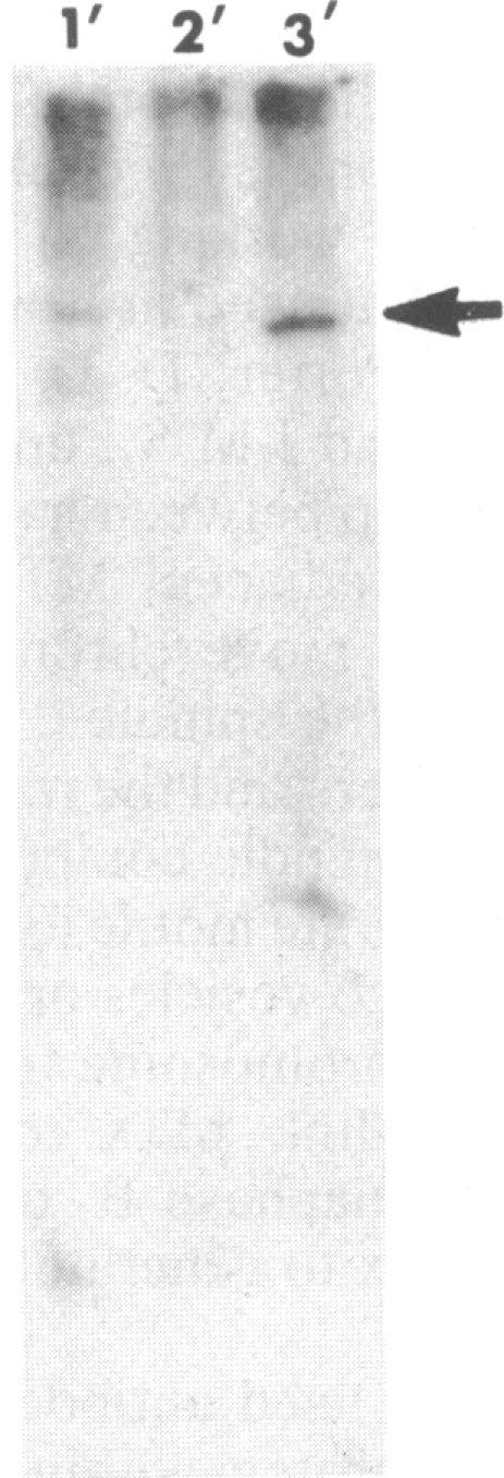
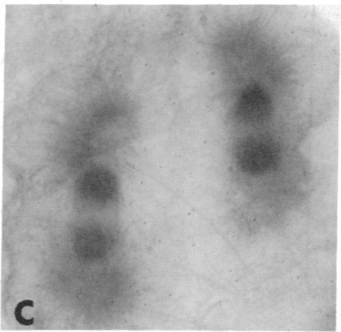
Sea urchin embryos in second division have been lysed into microtubule-stabilizing buffers to yield mitotic cytoskeletons (MCSs) that consist of two mitotic spindles surrounded by a cortical array of filaments. Microtubules have been completely extracted from MCSs by incubation at 0 degrees C with Ca2+-containing buffer. An antibody to the microtubule translocator kinesin stains the spindles in MCSs and in MCSs treated with 5 mM ATP and also stains spindle-remnants of the MCSs after the microtubules have been extracted. We conclude that kinesin binds to a nonmicrotubule component in the mitotic spindle. Based on these results, we present several models of kinesin function in the spindle.
处于第二次分裂期的海胆胚胎已被裂解到微管稳定缓冲液中,以产生有丝分裂细胞骨架(MCS),其由两个被丝状皮质阵列包围的有丝分裂纺锤体组成。通过在0℃下与含钙离子的缓冲液孵育,微管已从MCS中完全提取出来。一种针对微管转运蛋白驱动蛋白的抗体可对MCS以及用5 mM ATP处理的MCS中的纺锤体进行染色,并且在微管被提取后也可对MCS的纺锤体残余物进行染色。我们得出结论,驱动蛋白与有丝分裂纺锤体中的一种非微管成分结合。基于这些结果,我们提出了驱动蛋白在纺锤体中功能的几种模型。