Department of Vascular Biology, Institute of Development, Aging and Cancer, Tohoku University, Sendai, Japan.
Division of Gastroenterology, Tohoku University Graduate School of Medicine, Sendai, Japan.
Cancer Sci. 2019 Jul;110(7):2296-2308. doi: 10.1111/cas.14041. Epub 2019 Jun 18.
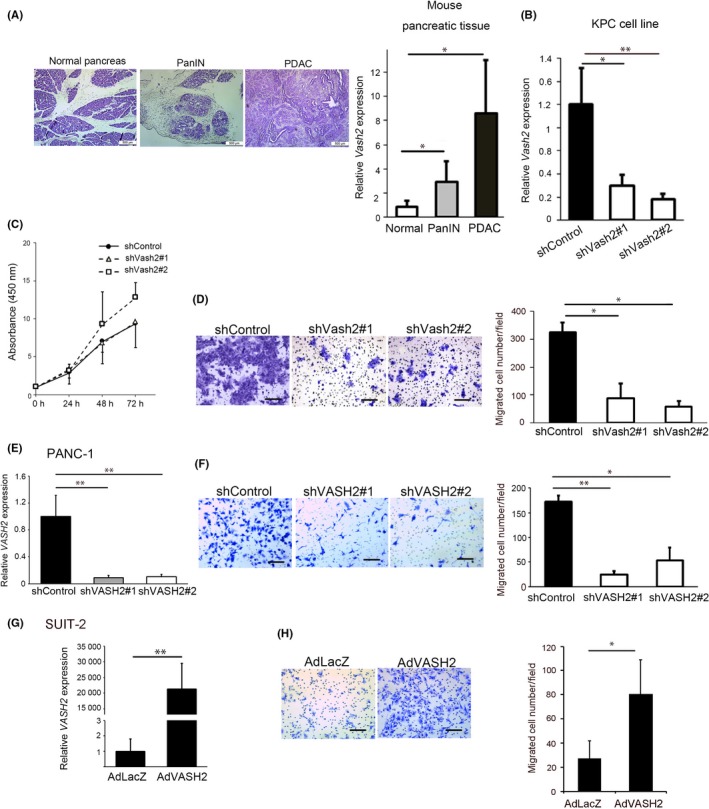
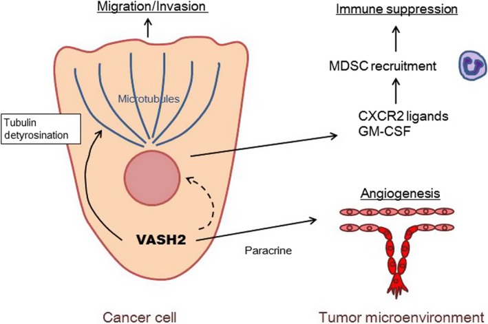
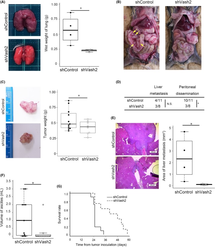
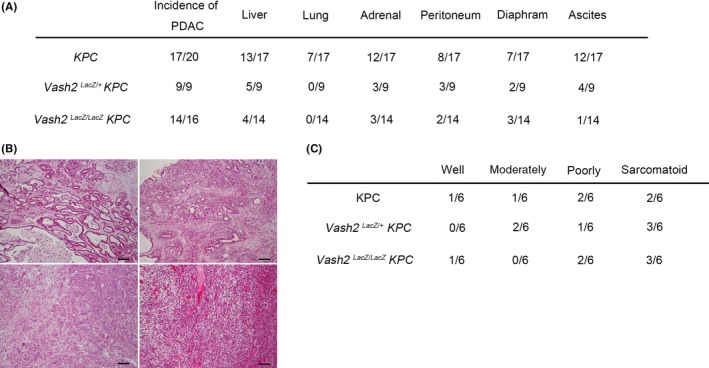
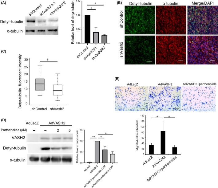
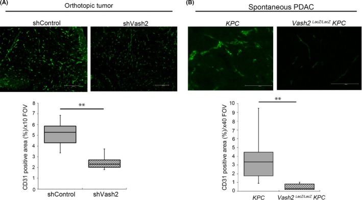
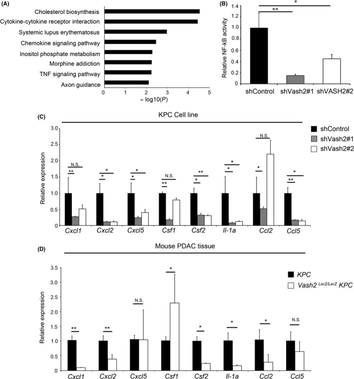
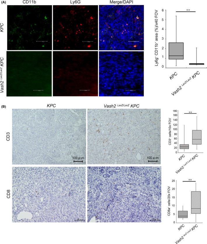
Vasohibin-2 (VASH2) is expressed in various cancers and promotes their progression. We recently reported that pancreatic cancer patients with higher VASH2 expression show poorer prognosis. Herein, we sought to characterize the role of VASH2 in pancreatic cancer. We used LSL-Kras ; LSL-Trp53 ; Pdx-1-Cre (KPC) mice, a mouse model of pancreatic ductal adenocarcinoma (PDAC), and cells isolated from them (KPC cells). Knockdown of Vash2 from PDAC cells did not affect their proliferation, but decreased their migration. When Vash2-knockdown PDAC cells were orthotopically inoculated, liver metastasis and peritoneal dissemination were reduced, and the survival period was significantly prolonged. When KPC mice were crossed with Vash2-deficient mice, metastasis was significantly decreased in Vash2-deficient KPC mice. VASH2 was recently identified to have tubulin carboxypeptidase activity. VASH2 knockdown decreased, whereas VASH2 overexpression increased tubulin detyrosination of PDAC cells, and tubulin carboxypeptidase (TCP) inhibitor parthenolide inhibited VASH2-induced cell migration. We next clarified its role in the tumor microenvironment. Tumor angiogenesis was significantly abrogated in vivo when VASH2 was knocked down or deleted. We further examined genes downregulated by Vash2 knockdown in KPC cells, and found chemokines and cytokines that were responsible for the recruitment of myeloid derived suppressor cells (MDSC). Indeed, MDSC were accumulated in PDAC of KPC mice, and they were significantly decreased in Vash2-deficient KPC mice. These findings suggest that VASH2 plays an essential role in the metastasis of PDAC with multiple effects on both cancer cells and the tumor microenvironment, including tubulin detyrosination, tumor angiogenesis and evasion of tumor immunity.
血管生成抑制素 2(VASH2)在各种癌症中表达,并促进其进展。我们最近报道,VASH2 表达较高的胰腺癌患者预后较差。在此,我们试图研究 VASH2 在胰腺癌中的作用。我们使用 LSL-Kras;LSL-Trp53;Pdx-1-Cre(KPC)小鼠,一种胰腺导管腺癌(PDAC)的小鼠模型,以及从它们分离的细胞(KPC 细胞)。从 PDAC 细胞中敲低 Vash2 不会影响其增殖,但会降低其迁移。当 Vash2 敲低的 PDAC 细胞原位接种时,肝转移和腹膜扩散减少,生存时间显著延长。当 KPC 小鼠与 Vash2 缺陷小鼠杂交时,Vash2 缺陷的 KPC 小鼠的转移明显减少。VASH2 最近被鉴定具有微管羧肽酶活性。VASH2 敲低降低,而 VASH2 过表达增加 PDAC 细胞的微管脱酪氨酸化,微管羧肽酶(TCP)抑制剂白头翁素抑制 VASH2 诱导的细胞迁移。我们接下来阐明了它在肿瘤微环境中的作用。体内敲低或缺失 VASH2 时,肿瘤血管生成显著受到抑制。我们进一步研究了 KPC 细胞中 Vash2 敲低下调的基因,发现了负责招募髓源性抑制细胞(MDSC)的趋化因子和细胞因子。事实上,KPC 小鼠的 PDAC 中 MDSC 积累,而 Vash2 缺陷的 KPC 小鼠中 MDSC 明显减少。这些发现表明 VASH2 在 PDAC 的转移中发挥着重要作用,对癌细胞和肿瘤微环境都有多种影响,包括微管脱酪氨酸化、肿瘤血管生成和逃避肿瘤免疫。