Pairaiturkar Pradyumna Purushottam, Sudame Onkar Shekhar, Pophale Chetan Shashikant
Department of Spine, Deenanath Mangeshkar Hospital, Pune, India.
J Korean Neurosurg Soc. 2019 Jul;62(4):414-421. doi: 10.3340/jkns.2018.0091. Epub 2019 May 14.
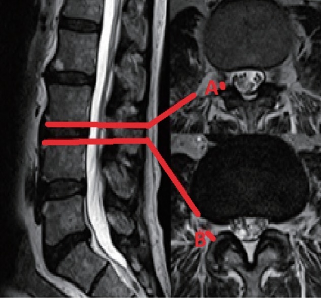
To evaluate 3-dimensional magnetic resonance imaging (MRI) of Kambin's safe zone to calculate maximum cannula diameter permissible for safe percutaneous endoscopic lumbar discectomy.
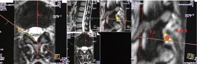
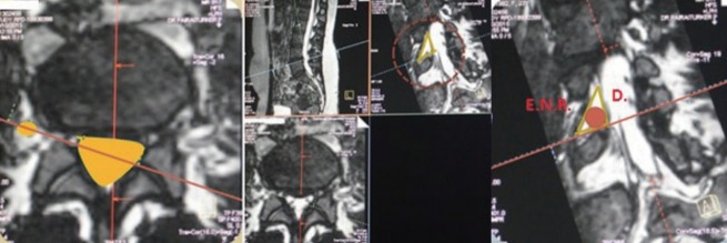
Fifty 3D MRIs of 19 males and 31 females (mean, 47 years) were analysed. Oblique, axial and sagittal views were used for image analysis. Three authors calculated the inscribed circle (cannula diameter) individually, within the neural (original) and bony Kambin's triangle in oblique views, disc heights on sagittal views and root to facet distances at upper and lower end plate levels on axial views and their averages were taken.
The mean root to facet distances at upper end plate level measured on axial sections increased from 3.42±3.01 mm at L12 level to 4.57±2.49 mm at L5S1 level. The mean root to facet distances at lower end plate level measured on axial sections also increased from 6.07±1.13 mm at L12 level to 12.9±2.83 mm at L5S1 level. Mean maximum cannula diameter permissible through the neural Kambin's triangle increased from 5.67±1.38 mm at L12 level to 9.7±3.82 mm at L5S1 level. The mean maximum cannula diameter permissible through the bony Kambin's triangle also increased from 4.03±1.08 mm at L12 level to 6.11±1 mm at L5S1 level. Only 2% of the 427 bony Kambin's triangles could accommodate a cannula diameter of 8mm. The base of the bony Kambin's triangle taken in oblique view (3D MRI) was significantly higher than the root to facet distance at lower end plate level taken in axial view.
The largest mean diameter of endoscopic cannula passable through "bony" Kambin's triangle was distinctively smaller than the largest mean diameter of endoscopic cannula passable through "neural" Kambin's triangle at all levels. Although proximity of exiting root to the facet joint is always taken into consideration before PELD procedure, our 3D MRI based anatomical study is the first to provide actual maximum cannula dimensions permissible in this region.
评估坎宾安全区的三维磁共振成像(MRI),以计算安全经皮内镜下腰椎间盘切除术允许的最大套管直径。
分析了19名男性和31名女性(平均47岁)的50例三维MRI。使用斜位、轴位和矢状位视图进行图像分析。三位作者分别计算斜位视图中神经(原始)和骨性坎宾三角内的内切圆(套管直径)、矢状位视图中的椎间盘高度以及轴位视图中上、下终板水平处神经根至关节突的距离,并取其平均值。
轴位切片上测量的上终板水平处神经根至关节突的平均距离从L12水平的3.42±3.01mm增加到L5S1水平的4.57±2.49mm。轴位切片上测量的下终板水平处神经根至关节突的平均距离也从L12水平的6.07±1.13mm增加到L5S1水平的12.9±2.83mm。经神经坎宾三角允许的平均最大套管直径从L12水平的5.67±1.38mm增加到L5S1水平的9.7±3.82mm。经骨性坎宾三角允许的平均最大套管直径也从L12水平的4.03±1.08mm增加到L5S1水平的6.11±1mm。在427个骨性坎宾三角中,只有2%能够容纳直径为8mm的套管。斜位视图(三维MRI)中骨性坎宾三角的底边明显高于轴位视图中下终板水平处神经根至关节突的距离。
在所有节段,经“骨性”坎宾三角可通过的内镜套管的最大平均直径明显小于经“神经”坎宾三角可通过的内镜套管的最大平均直径。尽管在经皮内镜下腰椎间盘切除术(PELD)手术前总是会考虑出口神经根与关节突关节的接近程度,但我们基于三维MRI的解剖学研究首次提供了该区域实际允许的最大套管尺寸。