Neurosurgery, National Neuroscience Institute, Singapore 308433, Singapore.
School of Electrical and Electronic Engineering, Nanyang Technological University, Singapore 639798, Singapore.
Sensors (Basel). 2019 May 10;19(9):2167. doi: 10.3390/s19092167.
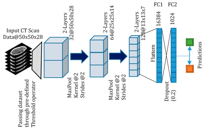
Intracranial hemorrhage is a medical emergency that requires urgent diagnosis and immediate treatment to improve patient outcome. Machine learning algorithms can be used to perform medical image classification and assist clinicians in diagnosing radiological scans. In this paper, we apply 3-dimensional convolutional neural networks (3D CNN) to classify computed tomography (CT) brain scans into normal scans (N) and abnormal scans containing subarachnoid hemorrhage (SAH), intraparenchymal hemorrhage (IPH), acute subdural hemorrhage (ASDH) and brain polytrauma hemorrhage (BPH). The dataset used consists of 399 volumetric CT brain images representing approximately 12,000 images from the National Neuroscience Institute, Singapore. We used a 3D CNN to perform both 2-class (normal versus a specific abnormal class) and 4-class classification (between normal, SAH, IPH, ASDH). We apply image thresholding at the image pre-processing step, that improves 3D CNN classification accuracy and performance by accentuating the pixel intensities that contribute most to feature discrimination. For 2-class classification, the F1 scores for various pairs of medical diagnoses ranged from 0.706 to 0.902 without thresholding. With thresholding implemented, the F1 scores improved and ranged from 0.919 to 0.952. Our results are comparable to, and in some cases, exceed the results published in other work applying 3D CNN to CT or magnetic resonance imaging (MRI) brain scan classification. This work represents a direct application of a 3D CNN to a real hospital scenario involving a medically emergent CT brain diagnosis.
颅内出血是一种医疗急症,需要紧急诊断和立即治疗,以改善患者的预后。机器学习算法可用于进行医学图像分类,并协助临床医生诊断放射扫描。在本文中,我们应用三维卷积神经网络(3D CNN)将计算机断层扫描(CT)脑扫描分为正常扫描(N)和异常扫描,异常扫描包含蛛网膜下腔出血(SAH)、脑实质内出血(IPH)、急性硬膜下血肿(ASDH)和脑多发创伤性出血(BPH)。使用的数据集由 399 个容积 CT 脑图像组成,代表来自新加坡国家神经科学研究所的大约 12000 个图像。我们使用 3D CNN 进行 2 类(正常与特定异常类)和 4 类(正常、SAH、IPH、ASDH 之间)分类。我们在图像预处理步骤中应用图像阈值处理,通过突出对特征区分贡献最大的像素强度,来提高 3D CNN 的分类准确性和性能。对于 2 类分类,在没有阈值的情况下,各种医学诊断对的 F1 分数范围为 0.706 至 0.902。实施阈值后,F1 分数提高,范围为 0.919 至 0.952。我们的结果与应用 3D CNN 进行 CT 或磁共振成像(MRI)脑扫描分类的其他工作的结果相当,在某些情况下甚至超过了这些结果。这项工作代表了将 3D CNN 直接应用于涉及医疗紧急 CT 脑诊断的真实医院场景。