Institute of Biomedical and Clinical Sciences, University of Exeter Medical School, Barrack Road, Exeter EX2 5DW, UK.
Wolfson Centre for Age-Related Diseases, King's College London, London WC2R 2LS, UK.
Hum Mol Genet. 2019 Aug 15;28(16):2763-2774. doi: 10.1093/hmg/ddz094.
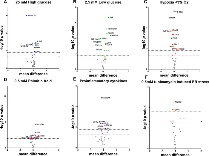
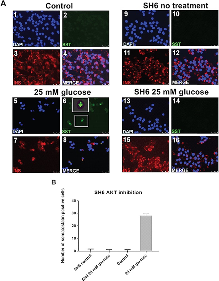
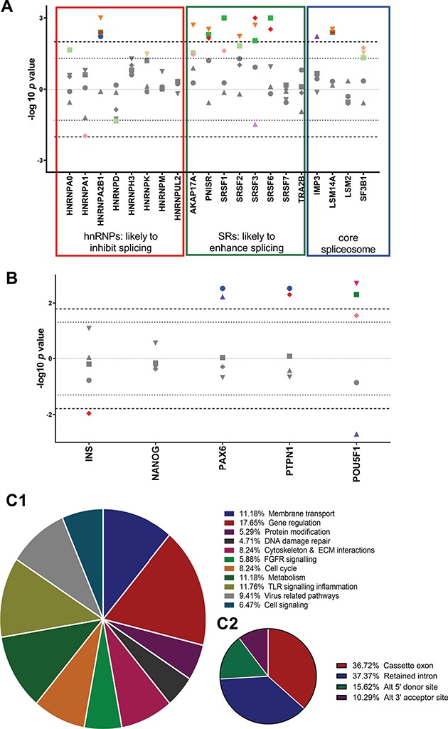
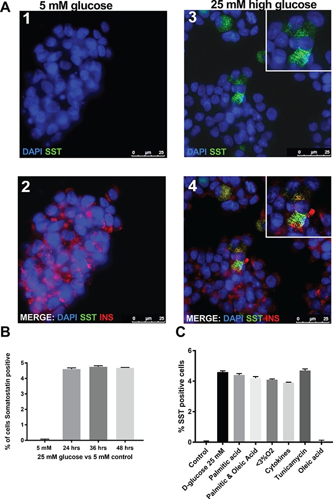
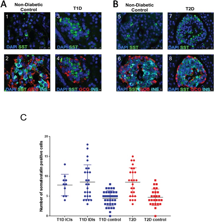
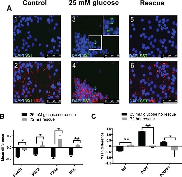
Changes to islet cell identity in response to type 2 diabetes (T2D) have been reported in rodent models, but are less well characterized in humans. We assessed the effects of aspects of the diabetic microenvironment on hormone staining, total gene expression, splicing regulation and the alternative splicing patterns of key genes in EndoC-βH1 human beta cells. Genes encoding islet hormones [somatostatin (SST), insulin (INS), Glucagon (GCG)], differentiation markers [Forkhead box O1 (FOXO1), Paired box 6, SRY box 9, NK6 Homeobox 1, NK6 Homeobox 2] and cell stress markers (DNA damage inducible transcript 3, FOXO1) were dysregulated in stressed EndoC-βH1 cells, as were some serine arginine rich splicing factor splicing activator and heterogeneous ribonucleoprotein particle inhibitor genes. Whole transcriptome analysis of primary T2D islets and matched controls demonstrated dysregulated splicing for ~25% of splicing events, of which genes themselves involved in messenger ribonucleic acid processing and regulation of gene expression comprised the largest group. Approximately 5% of EndoC-βH1 cells exposed to these factors gained SST positivity in vitro. An increased area of SST staining was also observed ex vivo in pancreas sections recovered at autopsy from donors with type 1 diabetes (T1D) or T2D (9.3% for T1D and 3% for T2D, respectively compared with 1% in controls). Removal of the stressful stimulus or treatment with the AKT Serine/Threonine kinase inhibitor SH-6 restored splicing factor expression and reversed both hormone staining effects and patterns of gene expression. This suggests that reversible changes in hormone expression may occur during exposure to diabetomimetic cellular stressors, which may be mediated by changes in splicing regulation.
已经有报道称,2 型糖尿病(T2D)会引起胰岛细胞的表型改变,但其在人类中的特征尚不明确。我们评估了糖尿病微环境的多个方面对 EndoC-βH1 人胰岛β细胞中激素染色、总基因表达、剪接调控以及关键基因的可变剪接模式的影响。在应激的 EndoC-βH1 细胞中,编码胰岛激素(生长抑素[SST]、胰岛素[INS]、胰高血糖素[GCG])、分化标志物(叉头框蛋白 O1[FOXO1]、配对盒基因 6[PAX6]、SRY 盒基因 9[SOX9]、NK6 同源盒 1[NKX6.1]、NK6 同源盒 2[NKX6.2])和细胞应激标志物(DNA 损伤诱导转录物 3[DDIT3]、FOXO1)的基因发生失调,一些丝氨酸/精氨酸丰富剪接因子剪接激活因子和异质核糖核蛋白颗粒抑制剂基因也是如此。对原发性 T2D 胰岛和匹配对照的全转录组分析表明,约 25%的剪接事件发生剪接失调,其中涉及信使核糖核酸处理和基因表达调控的基因本身构成了最大的组。大约 5%的暴露于这些因素的 EndoC-βH1 细胞在体外获得 SST 阳性。在从患有 1 型糖尿病(T1D)或 2 型糖尿病(T2D)的供体中获得的胰腺切片的离体研究中也观察到 SST 染色面积增加(与对照组的 1%相比,T1D 为 9.3%,T2D 为 3%)。去除应激刺激或用 AKT 丝氨酸/苏氨酸激酶抑制剂 SH-6 治疗可恢复剪接因子的表达,并逆转激素染色和基因表达模式的变化。这表明,在暴露于糖尿病样细胞应激时,激素表达可能发生可逆变化,这可能是由剪接调控的变化介导的。