He Jun, Yang Zhulin, Wu Zhengchun, Wang Lingxiang, Xu Shu, Zou Qiong, Yuan Yuan, Li Daiqiang
Hunan Provincial Key Laboratory of Hepatobiliary Disease Research, Department of General Surgery, Second Xiangya Hospital, Central South University, Changsha, Hunan, People's Republic of China.
Department of Pathology, Third Xiangya Hospital, Central South University, Changsha, Hunan, People's Republic of China.
Onco Targets Ther. 2019 Apr 17;12:2955-2965. doi: 10.2147/OTT.S197001. eCollection 2019.
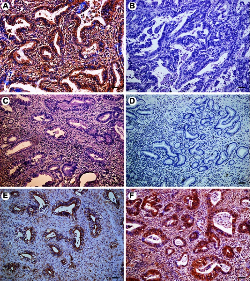
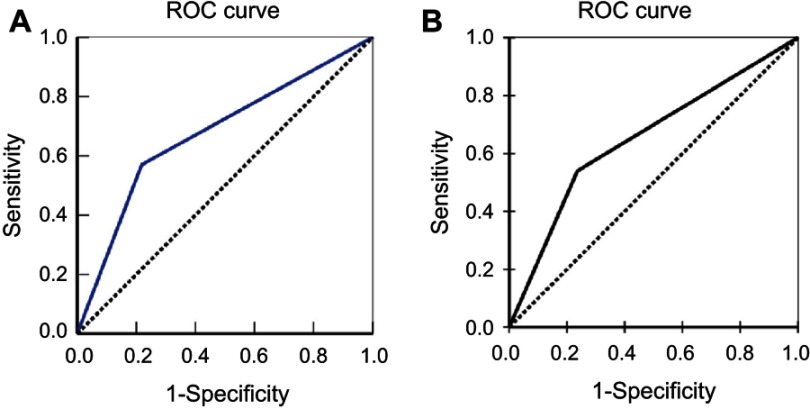
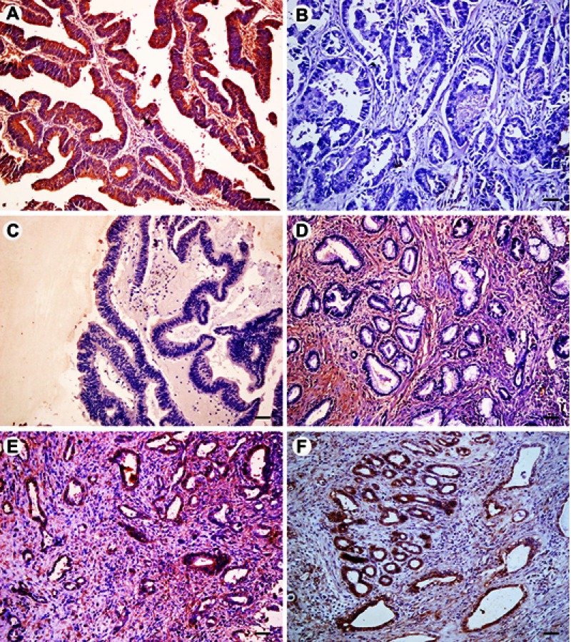
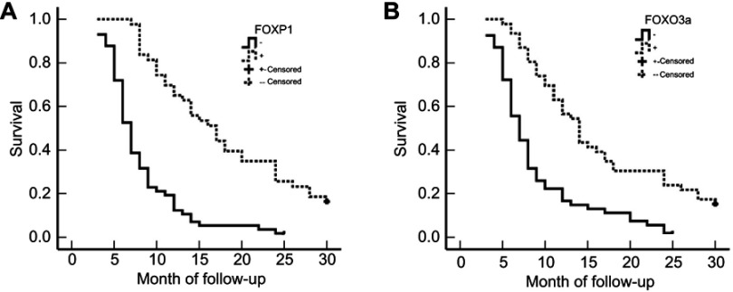
Extrahepatic cholangiocarcinoma (EHCC) is a highly malignant tumor with poor prognosis and intrinsic resistance to cytotoxic agents. The molecular mechanisms associated with high malignancy and resistance to chemotherapy and radiotherapy have not been fully elucidated. This study investigated the clinicopathological significances of FOXP1 and FOXO3a expression in EHCC. We assayed FOXP1 and FOXO3a expressions in 100 EHCC, 30 peritumoral tissues, 10 adenoma and 15 normal biliary tract tissues using EnVision immunohistochemistry. The positive rates of FOXP1 and FOXO3a proteins were significantly lower in EHCC tumors than in peritumoral tissues, adenoma, and normal bile tract tissues (<0.05 or <0.01). Adenoma and pericancerous tissues with negative FOXP1 and/or FOXO3a protein expressions exhibited atypical hyperplasia. The positive correlation was established between the expression of FOXP1 and FOXO3a in EHCC (<0.01). The positive rates of FOXP1 and FOXO3a expression were significantly higher in cases with well differentiation, no metastasis in lymph node, no invasion to surrounding tissues and organs, TNM I + II stage and radical resection (<0.05 or <0.01). Kaplan-Meier survival analysis showed that EHCC patients with positive FOXP1 and FOXO3a expression survived significantly higher than patients with negative FOXP1 and FOXO3a expression, respectively (<0.001). Cox multivariate analysis revealed that negative FOXP1 or FOXO3a expressions were independent poor prognostic factors in EHCC patients. The AUCs for FOXP1 and FOXO3a were 0.676 (95% CI: 0.589-0.763, <0.001) and 0.652 (95% CI: 0.563-741, =0.002), respectively. The present study indicates that negative FOXP1 and FOXO3a expressions are closely associated with the pathogenesis, clinical, pathological and biological behaviors, and poor prognosis in EHCC.
肝外胆管癌(EHCC)是一种预后较差且对细胞毒性药物具有内在抗性的高恶性肿瘤。与高恶性以及化疗和放疗抗性相关的分子机制尚未完全阐明。本研究调查了FOXP1和FOXO3a在EHCC中的表达的临床病理意义。我们使用EnVision免疫组织化学方法检测了100例EHCC、30例癌旁组织、10例腺瘤和15例正常胆道组织中FOXP1和FOXO3a的表达。EHCC肿瘤中FOXP1和FOXO3a蛋白的阳性率显著低于癌旁组织、腺瘤和正常胆道组织(<0.05或<0.01)。FOXP1和/或FOXO3a蛋白表达阴性的腺瘤和癌旁组织表现出非典型增生。EHCC中FOXP1和FOXO3a的表达之间存在正相关(<0.01)。在高分化、无淋巴结转移、未侵犯周围组织和器官、TNM I + II期以及根治性切除的病例中,FOXP1和FOXO3a表达的阳性率显著更高(<0.05或<0.01)。Kaplan-Meier生存分析表明,FOXP1和FOXO3a表达阳性的EHCC患者的生存率分别显著高于FOXP1和FOXO3a表达阴性的患者(<0.001)。Cox多因素分析显示,FOXP1或FOXO3a表达阴性是EHCC患者独立的不良预后因素。FOXP1和FOXO3a的曲线下面积分别为0.676(95%CI:0.589 - 0.763,<0.001)和0.652(95%CI:0.563 - 741,=0.002)。本研究表明,FOXP1和FOXO3a表达阴性与EHCC的发病机制、临床、病理和生物学行为以及不良预后密切相关。