Kum Su Jung, Lee Hye Won, Jung Hye Ra, Choe Misun, Kim Sang Pyo
Department of Pathology, Keimyung University School of Medicine, Daegu, Korea.
J Pathol Transl Med. 2019 Sep;53(5):327-331. doi: 10.4132/jptm.2019.05.14. Epub 2019 May 24.
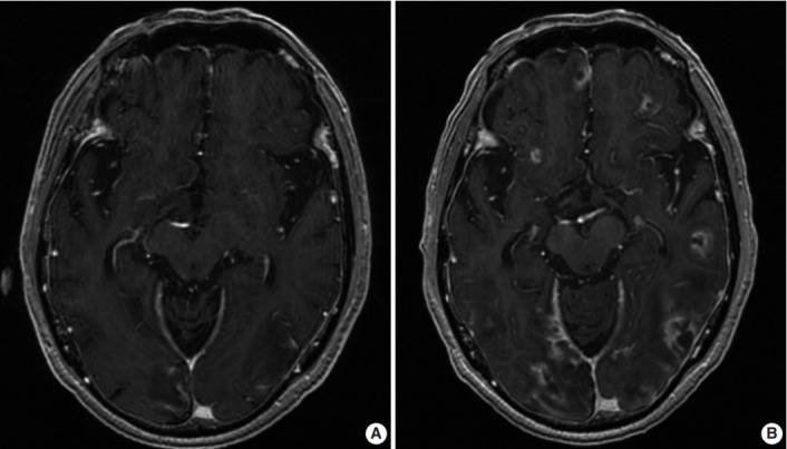
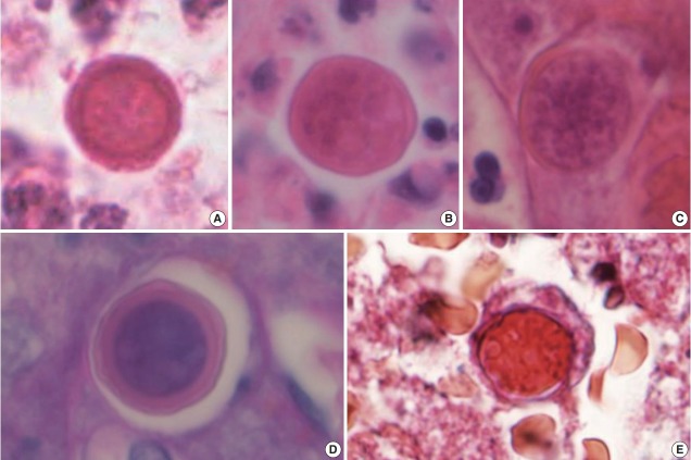
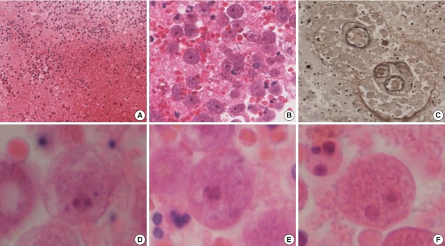
We present the case of a 71-year-old man who was diagnosed with amoebic encephalitis caused by Balamuthia mandrillaris. He had rheumatic arthritis for 30 years and had undergone continuous treatment with immunosuppressants. First, he complained of partial spasm from the left thigh to the left upper limb. Magnetic resonance imaging revealed multifocal enhancing nodules in the cortical and subcortical area of both cerebral hemispheres, which were suggestive of brain metastases. However, the patient developed fever with stuporous mentality and an open biopsy was performed immediately. Microscopically, numerous amoebic trophozoites, measuring 20 to 25 µm in size, with nuclei containing one to four nucleoli and some scattered cysts having a double-layered wall were noted in the background of hemorrhagic necrosis. Based on the microscopic findings, amoebic encephalitis caused by Balamuthia mandrillaris was diagnosed. The patient died on the 10th day after being admitted at the hospital. The diagnosis of amoebic encephalitis in the early stage is difficult for clinicians. Moreover, most cases undergo rapid deterioration, resulting in fatal consequences. In this report, we present the first case of B. mandrillaris amoebic encephalitis with fatal progression in a Korean patient.
我们报告一例71岁男性,其被诊断为由曼氏巴通体引起的阿米巴性脑炎。他患有风湿性关节炎30年,一直在接受免疫抑制剂持续治疗。起初,他主诉从左大腿至左上肢部分痉挛。磁共振成像显示双侧大脑半球皮质和皮质下区域有多发强化结节,提示脑转移瘤。然而,患者出现发热伴神志不清,随即立即进行了开放性活检。显微镜下可见,在出血性坏死背景中,有大量阿米巴滋养体,大小为20至25微米,细胞核含一至四个核仁,还有一些散在的双层壁囊肿。根据显微镜检查结果,诊断为由曼氏巴通体引起的阿米巴性脑炎。患者入院第10天死亡。临床医生早期诊断阿米巴性脑炎较为困难。此外,大多数病例病情迅速恶化,导致致命后果。在本报告中,我们呈现了首例韩国患者因曼氏巴通体引起的阿米巴性脑炎且病情呈致命进展的病例。