Solomon H. Snyder Department of Neuroscience, Johns Hopkins University School of Medicine, Baltimore, MD 21205, USA.
Solomon H. Snyder Department of Neuroscience, Johns Hopkins University School of Medicine, Baltimore, MD 21205, USA; Department of Oncology, Division of Biostatistics and Bioinformatics, Sidney Kimmel Comprehensive Cancer Center, Johns Hopkins University School of Medicine, Baltimore, MD 21205, USA; McKusick-Nathans Institute for Genetic Medicine, Johns Hopkins University School of Medicine, Baltimore, MD 21205, USA; Institute for Data Intensive Engineering and Science, Johns Hopkins University School of Medicine, Baltimore, MD 21205, USA.
Neuron. 2019 Jun 19;102(6):1111-1126.e5. doi: 10.1016/j.neuron.2019.04.010. Epub 2019 May 22.
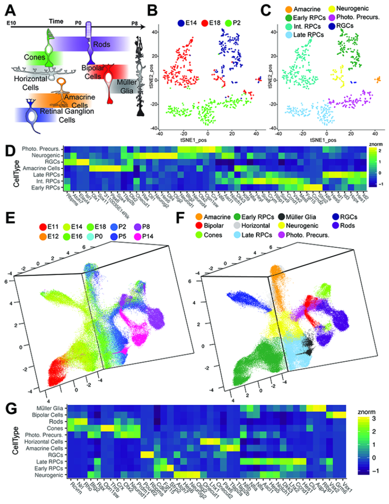
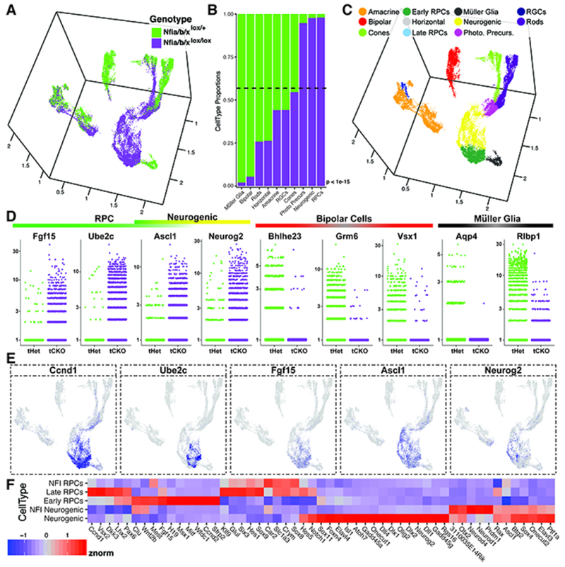
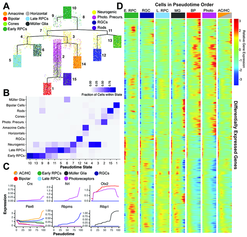
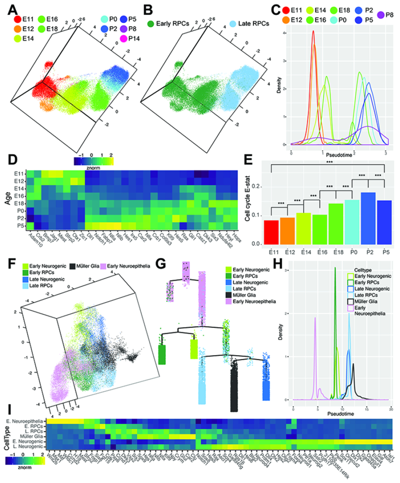
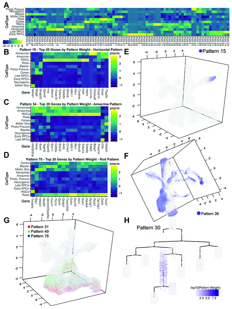
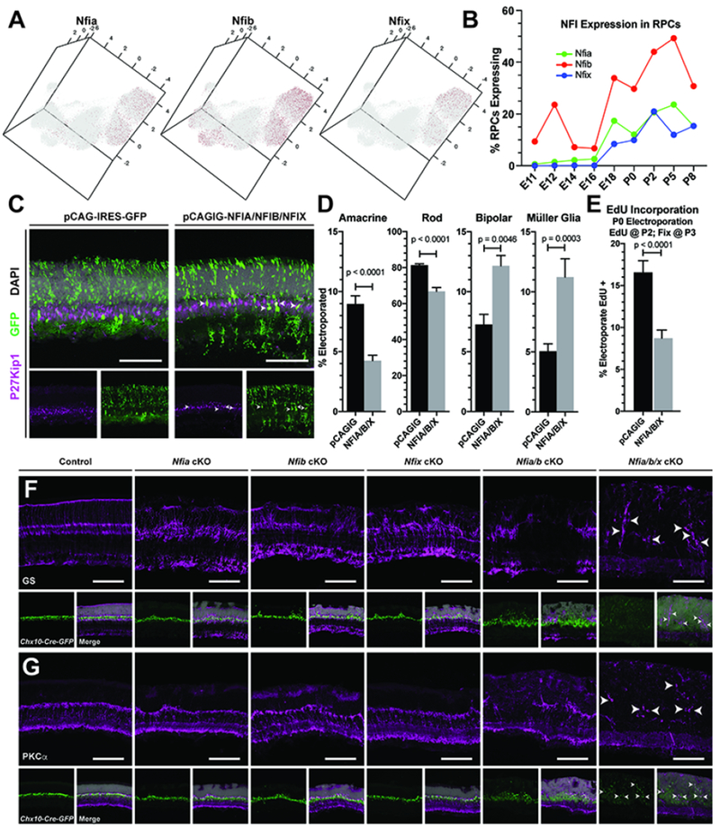
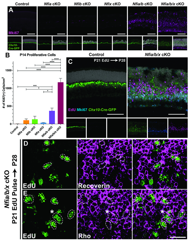
Precise temporal control of gene expression in neuronal progenitors is necessary for correct regulation of neurogenesis and cell fate specification. However, the cellular heterogeneity of the developing CNS has posed a major obstacle to identifying the gene regulatory networks that control these processes. To address this, we used single-cell RNA sequencing to profile ten developmental stages encompassing the full course of retinal neurogenesis. This allowed us to comprehensively characterize changes in gene expression that occur during initiation of neurogenesis, changes in developmental competence, and specification and differentiation of each major retinal cell type. We identify the NFI transcription factors (Nfia, Nfib, and Nfix) as selectively expressed in late retinal progenitor cells and show that they control bipolar interneuron and Müller glia cell fate specification and promote proliferative quiescence.
精确控制神经祖细胞中的基因表达对于正确调节神经发生和细胞命运特化是必要的。然而,发育中中枢神经系统的细胞异质性一直是确定控制这些过程的基因调控网络的主要障碍。为了解决这个问题,我们使用单细胞 RNA 测序来描述包括视网膜神经发生全过程的十个发育阶段。这使我们能够全面描述神经发生起始时发生的基因表达变化、发育能力的变化以及每种主要视网膜细胞类型的特化和分化。我们确定 NFI 转录因子(Nfia、Nfib 和 Nfix)选择性地在晚期视网膜祖细胞中表达,并表明它们控制双极中间神经元和 Müller 胶质细胞命运特化,并促进增殖静止。