Institute of Medical Science, University of Toronto, Toronto, ON, Canada.
Department of Obstetrics and Gynecology, University of Toronto, Toronto, ON, Canada.
Theranostics. 2019 Apr 13;9(9):2727-2738. doi: 10.7150/thno.31225. eCollection 2019.
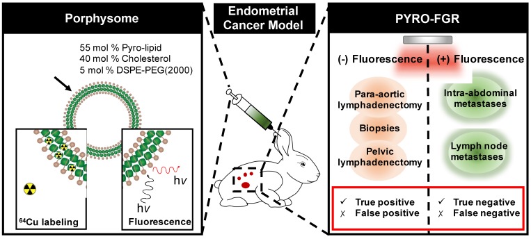
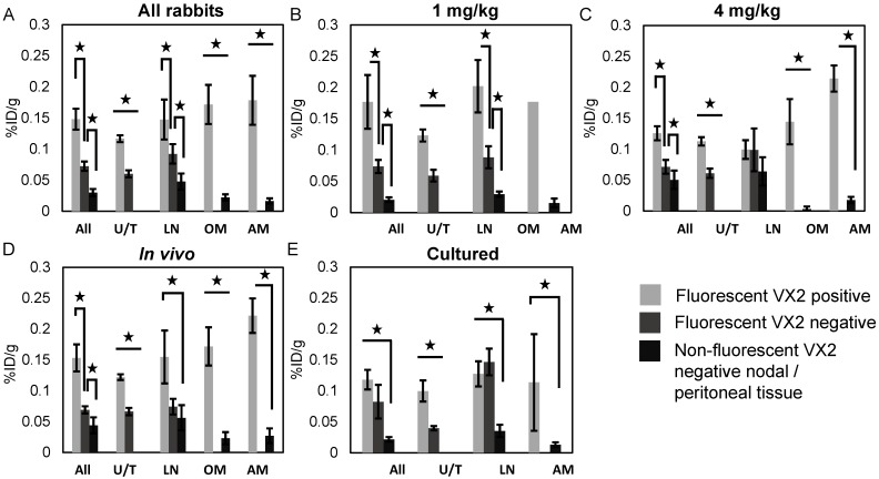
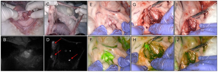
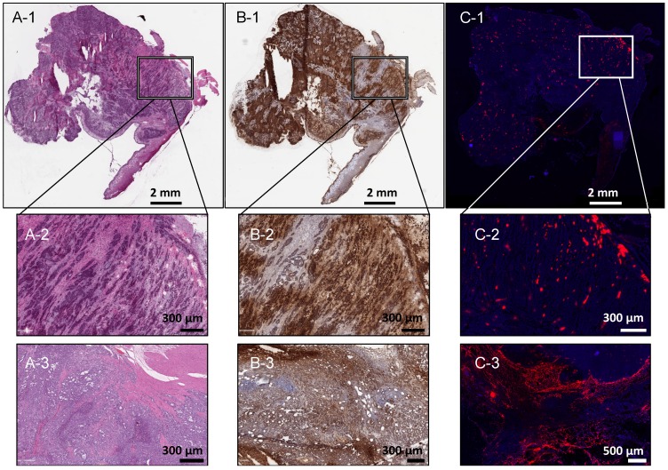
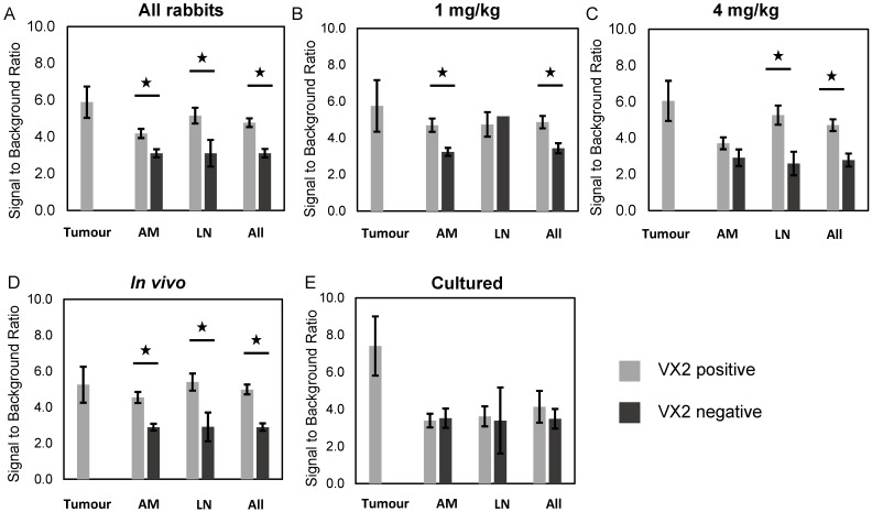
: To investigate Porphysome fluorescence image-guided resection (PYRO-FGR) for detection of uterine tumour, metastatic lymph nodes and abdominal metastases in a model of endometrial cancer. : White New Zealand rabbits were inoculated with VX2 cells via intra-myometrial injection. At 30 days, Porphysomes were administered intravenously. At 24 h the abdomen was imaged and fluorescent tissue identified (PYRO-FGR). After complete resection of fluorescent tissue, fluorescence-negative lymph nodes and peritoneal biopsies were removed. Histopathology including ultra-staging and analysis by a pathologist was used to detect tumour. Fluorescence signal to background ratio (SBR) was calculated and VX2 (+) tissue compared to VX2 (-) tissue. Biodistribution was calculated and Porphysome accumulation in fluorescent VX2 (+) tissue compared to fluorescent VX2 (-) and non-fluorescent VX2 (-) tissue. : Of 17 VX2 models, 10 received 4 mg/kg of Porphysomes and 7 received 1 mg/kg. Seventeen tumours (UT), 81 lymph nodes (LN) and 54 abdominal metastases (AM) were fluorescence-positive and resected. Of these, 17 UT, 60 LN and 45 AM were VX2 (+), while 16 LN and 5 AM were VX2 (-). Nine specimens were excluded from analysis. Thirty-one LN and 53 peritoneal biopsies were fluorescence-negative and resected. Of these, all LN and 51/53 biopsies were VX2 (-) with only 2 false-negative biopsies. Sensitivity and specificity of PYRO-FGR for VX2 (+) tissue was 98.4% / 80.0% overall, 100% / 100% for UT, 100% / 66.0 % for LN and 95.7% / 91.4% for AM. Increased SBR and biodistribution was observed in VX2 (+) tissue vs. VX2 (-) tissue. : Porphysomes are a highly sensitive imaging agent for intra-operative detection and resection of uterine tumour, metastatic lymph nodes and abdominal metastases.
: 研究卟啉体荧光图像引导切除(PYRO-FGR)在子宫内膜癌模型中检测子宫肿瘤、转移性淋巴结和腹部转移。: 通过子宫内注射向新西兰白兔接种 VX2 细胞。在第 30 天,静脉内给予卟啉体。在 24 小时时对腹部进行成像,并识别荧光组织(PYRO-FGR)。在完全切除荧光组织后,切除荧光阴性的淋巴结和腹膜活检。组织病理学检查包括超分期和病理学家分析,以检测肿瘤。计算荧光信号与背景比(SBR),并将 VX2(+)组织与 VX2(-)组织进行比较。计算生物分布,并将卟啉体在荧光 VX2(+)组织中的积累与荧光 VX2(-)和非荧光 VX2(-)组织进行比较。: 在 17 个 VX2 模型中,10 个接受 4mg/kg 的卟啉体,7 个接受 1mg/kg 的卟啉体。17 个肿瘤(UT)、81 个淋巴结(LN)和 54 个腹部转移(AM)呈荧光阳性并被切除。其中,17 个 UT、60 个 LN 和 45 个 AM 是 VX2(+),而 16 个 LN 和 5 个 AM 是 VX2(-)。有 9 个标本被排除在分析之外。31 个 LN 和 53 个腹膜活检呈荧光阴性并被切除。其中,所有 LN 和 51/53 个活检均为 VX2(-),仅 2 个活检为假阴性。PYRO-FGR 对 VX2(+)组织的敏感性和特异性总体为 98.4%/80.0%,对 UT 为 100%/100%,对 LN 为 100%/66.0%,对 AM 为 95.7%/91.4%。在 VX2(+)组织中观察到 SBR 和生物分布增加与 VX2(-)组织相比。: 卟啉体是一种高度敏感的术中检测和切除子宫肿瘤、转移性淋巴结和腹部转移的成像剂。