Department of Surgery and Oncology, Graduate School of Medical Sciences, Kyushu University, 3-1-1 Maidashi, Fukuoka, 812-8582, Japan.
Department of Advanced Medical Initiatives, Graduate School of Medical Sciences, Kyushu University, Fukuoka, Japan.
J Exp Clin Cancer Res. 2019 May 27;38(1):221. doi: 10.1186/s13046-019-1226-8.
Extracellular signal-regulated kinases (ERKs) have been related to multiple cancers, including breast cancer, hepatocellular cancer, lung cancer and colorectal cancer. ERK1/2 inhibitor can suppress growth of KRAS-mutant pancreatic tumors by targeting cancer cell. However, no studies have shown the expression of ERK1/2 on pancreatic stromal and its effect on pancreatic cancer-stromal interaction.
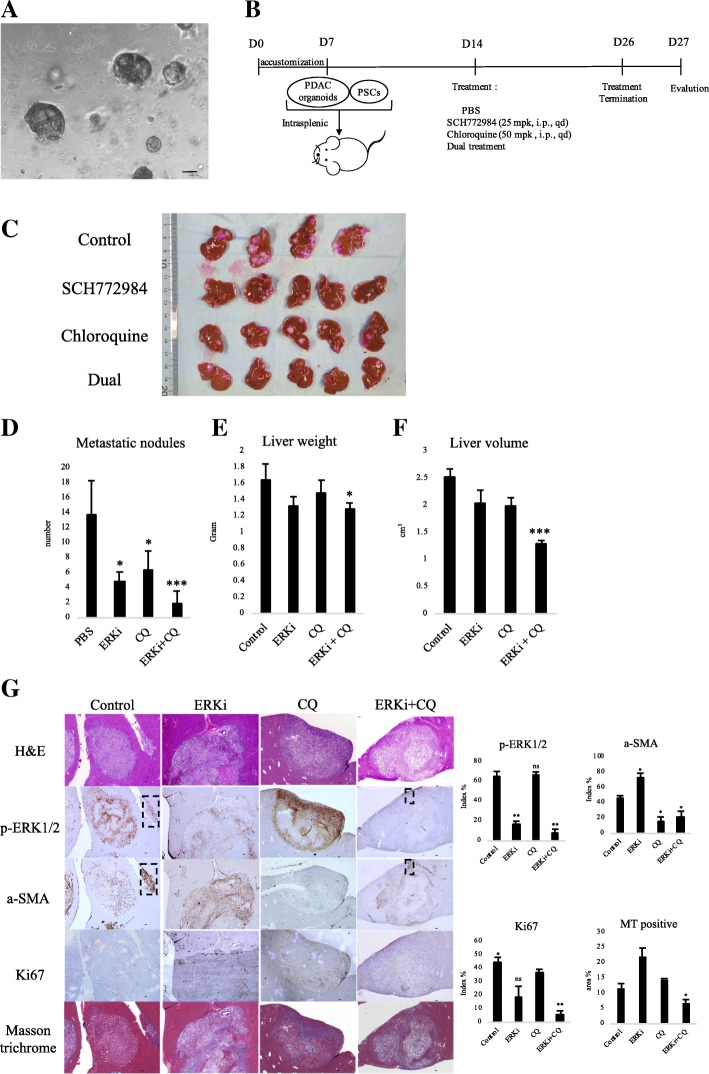
Immunohistochemistry and western blotting were performed to detect the expression of p-ERK1/2 in pancreatic tissues and cells. Cell viability assay was used to study IC50 of ERK inhibitor on pancreatic cancer cells (PCCs) and primary cancer-associated pancreatic stellate cells (PSCs). Transwell migration, invasion, cell viability assay, senescence β-galactosidase staining were performed to determine the effect of ERK inhibitor on PCCs and PSCs in vitro and in vivo. The expression of key factors involved in autophagy and epithelial-to-mesenchymal transition (EMT) process were evaluated by western blotting. The expression of key factors related to cell invasiveness and malignancy were confirmed by qRT-PCR. Co-transplantation of PCC Organoid and PSC using a splenic xenograft mouse model was used to evaluated combined treatment of ERK inhibitor and autophagy inhibitor.
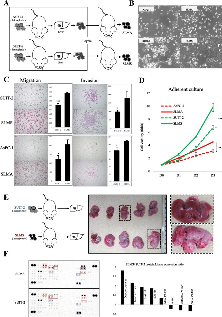
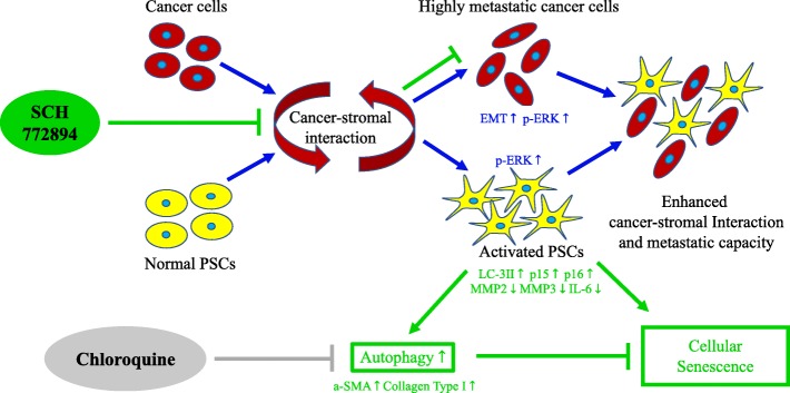
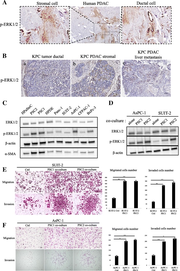
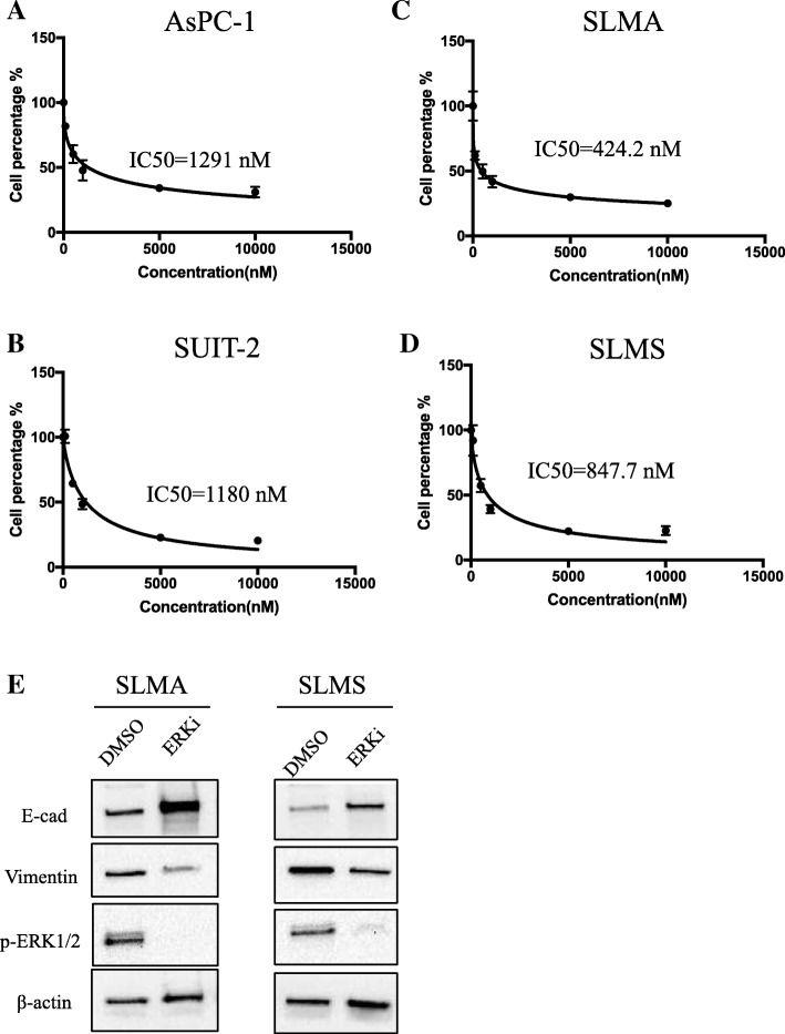
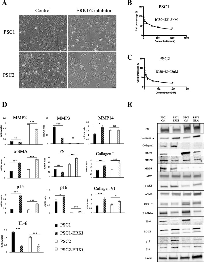
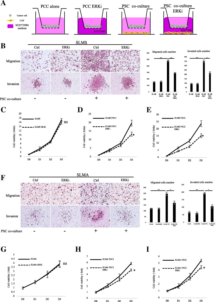
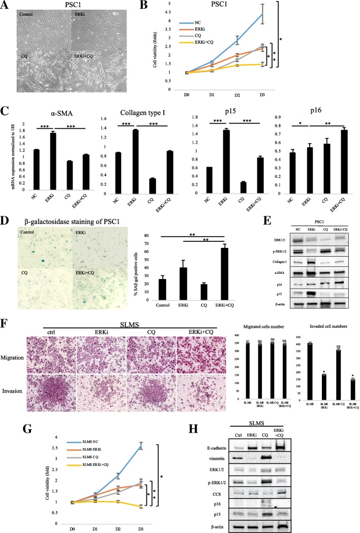
Immunohistochemical staining in pancreatic tumor samples and transgenetic mice detected p-ERK1/2 expression in both cancer cells and stromal cells. In pancreatic tissues, p-ERK1/2 was strongly expressed in cancer-associated PSCs compared with cancer cells and normal PSCs. PSCs were also significantly more sensitive to ERK1/2 inhibitor treatment. Inhibition of ERK1/2 suppressed EMT transition in HMPCCs, upregulated cellular senescence markers, activated autophagy in cancer-associated PSCs; and suppressed cancer-stromal interaction, which enhanced invasiveness and viability of cancer cells. We also found that chloroquine, an autophagy inhibitor, suppressed ERK inhibition-induced autophagy and promoted PSC cellular senescence, leading to significantly decreased cell proliferation. The combination of an ERK inhibitor and autophagy inhibitor suppressed liver metastasis in a splenic pancreatic cancer organoid xenograft mouse model.
These data indicate that inhibition of ERK1/2 in cancer-associated pancreatic stellate cells suppresses cancer-stromal interaction and metastasis.
细胞外信号调节激酶(ERK)与多种癌症有关,包括乳腺癌、肝细胞癌、肺癌和结直肠癌。ERK1/2 抑制剂通过靶向癌细胞可以抑制 KRAS 突变胰腺肿瘤的生长。然而,尚无研究表明 ERK1/2 在胰腺基质中的表达及其对胰腺癌-基质相互作用的影响。
采用免疫组织化学和 Western blot 检测胰腺组织和细胞中 p-ERK1/2 的表达。细胞活力测定法用于研究 ERK 抑制剂对胰腺癌细胞(PCCs)和原代癌相关胰腺星状细胞(PSCs)的 IC50。Transwell 迁移、侵袭、细胞活力测定、衰老β-半乳糖苷酶染色用于确定 ERK 抑制剂对体外和体内 PCCs 和 PSCs 的影响。通过 Western blot 评估自噬和上皮间质转化(EMT)过程中涉及的关键因子的表达。通过 qRT-PCR 证实与细胞侵袭性和恶性相关的关键因子的表达。使用脾异种移植小鼠模型进行 PCC 类器官和 PSC 的共移植,以评估 ERK 抑制剂和自噬抑制剂的联合治疗。
在胰腺肿瘤样本和转基因小鼠中进行免疫组织化学染色检测到 p-ERK1/2 在癌细胞和基质细胞中均有表达。在胰腺组织中,与癌细胞和正常 PSCs 相比,癌相关 PSCs 中 p-ERK1/2 表达强烈。PSCs 对 ERK1/2 抑制剂的治疗也更为敏感。ERK1/2 的抑制抑制了 HMPCCs 中的 EMT 转化,上调了细胞衰老标志物,激活了癌相关 PSCs 中的自噬,并抑制了癌-基质相互作用,从而增强了癌细胞的侵袭性和活力。我们还发现,自噬抑制剂氯喹抑制了 ERK 抑制诱导的自噬并促进了 PSC 细胞衰老,导致细胞增殖显著减少。ERK 抑制剂和自噬抑制剂的联合抑制了脾胰腺癌症类器官异种移植小鼠模型中的肝转移。
这些数据表明,抑制癌相关胰腺星状细胞中的 ERK1/2 可抑制癌-基质相互作用和转移。