Soheili-Nezhad Sourena, Sedghi Alireza, Schweser Ferdinand, Eslami Shahr Babaki Amir, Jahanshad Neda, Thompson Paul M, Beckmann Christian F, Sprooten Emma, Toghae Mansoureh
Donders Institute for Brain, Cognition, and Behaviour, Radboud University Medical Centre, Nijmegen, Netherlands.
Donders Centre for Cognitive Neuroimaging, Radboud University, Nijmegen, Netherlands.
Front Neurol. 2019 May 7;10:442. doi: 10.3389/fneur.2019.00442. eCollection 2019.
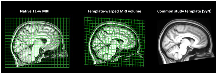
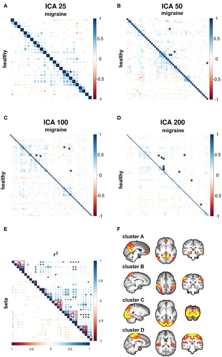
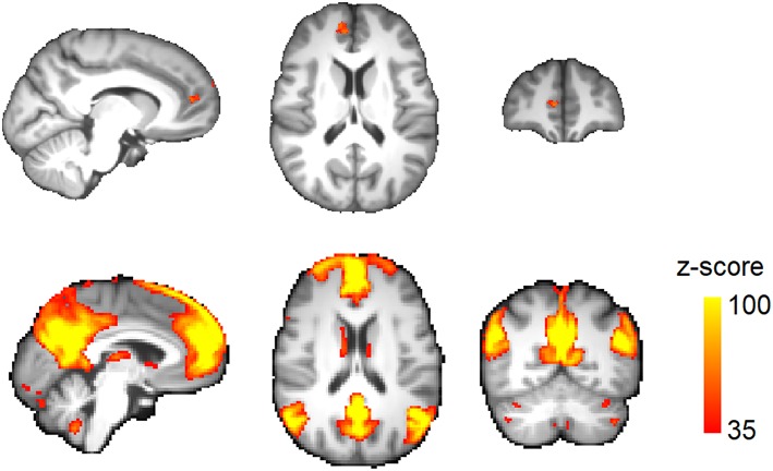
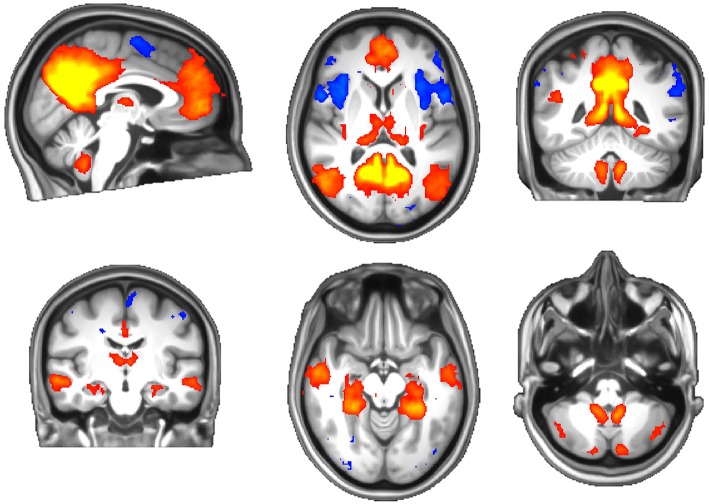
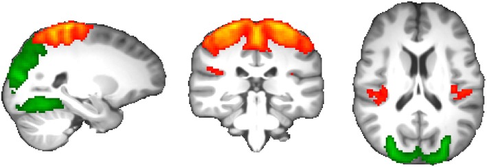
It remains unknown whether migraine headache has a progressive component in its pathophysiology. Quantitative MRI may provide valuable insight into abnormal changes in the migraine interictum and assist in identifying disrupted brain networks. We carried out a data-driven study of structural integrity and functional connectivity of the resting brain in migraine without aura. MRI scanning was performed in 36 patients suffering from episodic migraine without aura and 33 age-matched healthy subjects. Voxel-wise analysis of regional brain volume was performed by registration of the T1-weighted MRI scans into a common study brain template using the tensor-based morphometry (TBM) method. Changes in functional synchronicity of the brain networks were assessed using probabilistic independent component analysis (ICA). TBM revealed that migraine is associated with reduced volume of the medial prefrontal cortex (mPFC). Among 375 functional brain networks, resting-state connectivity was decreased between two components spanning the visual cortex, posterior insula, and parietal somatosensory cortex. Our study reveals structural and functional alterations of the brain in the migraine interictum that may stem from underlying disease risk factors and the "silent" aura phenomenon. Longitudinal studies will be needed to investigate whether interictal brain changes are progressive and associated with clinical disease trajectories.
偏头痛在其病理生理学中是否具有进行性成分仍不清楚。定量磁共振成像(MRI)可能为偏头痛发作间期的异常变化提供有价值的见解,并有助于识别受损的脑网络。我们对无先兆偏头痛患者静息态大脑的结构完整性和功能连接性进行了一项数据驱动的研究。对36例发作性无先兆偏头痛患者和33名年龄匹配的健康受试者进行了MRI扫描。使用基于张量的形态测量(TBM)方法,将T1加权MRI扫描图像配准到一个通用的研究脑模板中,对区域脑体积进行体素分析。使用概率独立成分分析(ICA)评估脑网络功能同步性的变化。TBM显示,偏头痛与内侧前额叶皮质(mPFC)体积减小有关。在375个功能性脑网络中,跨越视觉皮质、后岛叶和顶叶体感皮质的两个成分之间的静息态连接性降低。我们的研究揭示了偏头痛发作间期大脑的结构和功能改变,这些改变可能源于潜在的疾病危险因素和“沉默”的先兆现象。需要进行纵向研究,以调查发作间期脑变化是否具有进行性以及是否与临床疾病轨迹相关。