Private Dental Practice, Lipowa 18, Wschowa, Poland.
"Sapienza" University of Rome, Italy.
Biomed Res Int. 2019 Apr 22;2019:2785302. doi: 10.1155/2019/2785302. eCollection 2019.
Various procedures in dental implantology are performed to enhance the bone healing process and implant stability. One of these methods can be a low-level laser therapy (LLLT).
The aim of our study was to evaluate the stabilization (primary and secondary) and bone density in peri-implant zone after LLLT protocol using a 635 nm diode laser.
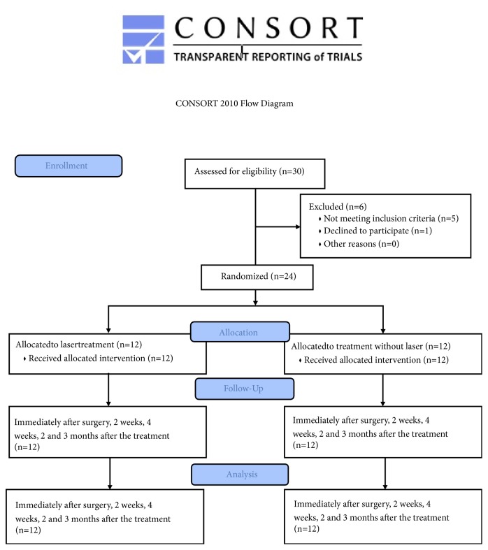
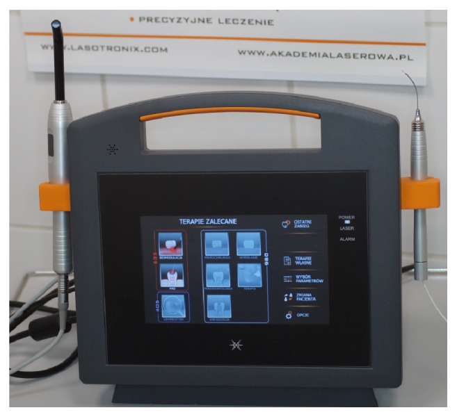
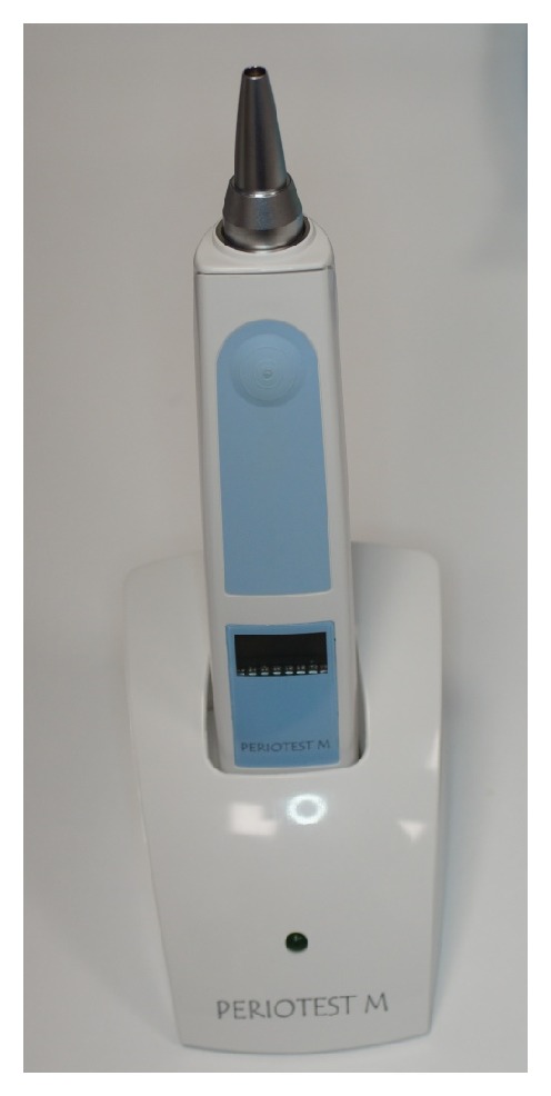
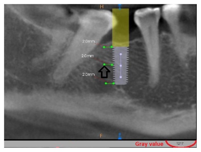
The research included 40 implants placed in the posterior region of a mandible in 24 patients (8 women and 16 man; age: 46.7 ± 8.7 years). The patients were randomly divided into 2 groups G1 (n=12, 18 implants) and G2 (n=12, 22 implants) according to the treatment procedure; G1 (test): 635 nm laser, with handpiece diameter: 8mm, output power: 100mW, spot area: 0.5024cm, average power density: 199.04mW/cm, continuous mode, dose: 4J per point (8J/cm), time: 40 sec per point, 2 points (irradiation on a buccal and a lingual side of the alveolus/implant), and total energy per session 8J; G2 (control): no laser irradiation. The G1 (test) group's implants were irradiated according to the following protocol: 1 day before surgery, immediately after the surgery and 2, 4, 7, and 14 days after. The total energy after all therapeutic sessions was 48J. The implants stability was measured employing a Periotest device (Periotest Test Value: PTV) (measured immediately after the surgery, 7 days, 2 weeks, 4 weeks, and 2 and 3 months after the surgery) and the bone density using cone-beam computed tomography (grayscale value) (measured immediately after the surgery, 4 weeks and 12 weeks after the treatment).
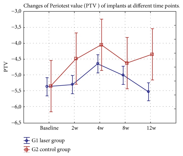
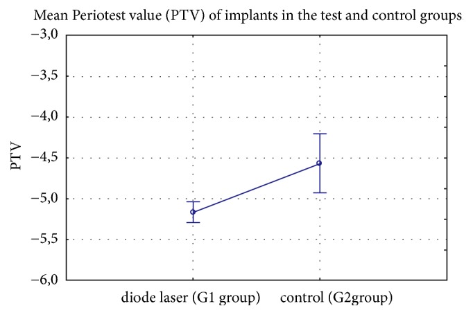
The average implant stability at different time points showed lower PTV value (higher stability) at 2 and 4 week after 635 nm laser irradiation (G1) compared with a control (G2) group (p<0.01). The secondary stability of the implants after 12 weeks observation was not significantly higher for the laser group in contrast to none-irradiated implants (p>0.05). The mean grayscale value at the apical, middle, and cervical level of the titanium implants showed the reduction of pixel grayscale value after 2 weeks and was lower for the G1 group in contrast to the G2 group (p<0.01). The value of grayscale after 12 weeks was significantly higher at the middle and apical level of the implants in the G1group in contrast to the G2 group (p<0.01).
The application of the 635 nm diode laser enhanced secondary implant stability and bone density. However, to assess the impact of the LLLT on peri-implant bone with different bone densities, further well-controlled long-term trials on larger study groups are needed.
在牙科种植体学中,各种程序被用于增强骨愈合过程和植入物稳定性。其中一种方法可以是低水平激光治疗(LLLT)。
我们的研究目的是评估使用 635nm 二极管激光的 LLLT 方案对种植体周围区的稳定(初级和次级)和骨密度的影响。
该研究包括在后牙区下颌骨中放置的 40 个种植体,涉及 24 名患者(8 名女性和 16 名男性;年龄:46.7±8.7 岁)。根据治疗程序,患者被随机分为两组 G1(n=12,18 个种植体)和 G2(n=12,22 个种植体);G1(试验):635nm 激光,手持件直径:8mm,输出功率:100mW,光斑面积:0.5024cm,平均功率密度:199.04mW/cm,连续模式,剂量:4J/点(8J/cm),时间:40 秒/点,2 点(牙槽/种植体颊侧和舌侧照射),每个疗程总能量 8J;G2(对照):无激光照射。G1(试验)组的种植体按照以下方案进行照射:手术前 1 天、手术后立即、术后 2、4、7 和 14 天。所有治疗疗程结束后的总能量为 48J。种植体稳定性使用 Periotest 设备(Periotest 测试值:PTV)(术后即刻、7 天、2 周、4 周和术后 2 个月和 3 个月测量)和骨密度使用锥形束计算机断层扫描(灰度值)(术后即刻、4 周和治疗后 12 周测量)。
不同时间点的平均种植体稳定性显示,与对照组(G2)相比,635nm 激光照射后 2 周和 4 周时 PTV 值(稳定性更高)较低(p<0.01)。与未照射的种植体相比,激光组在 12 周观察期的次级稳定性没有显著提高(p>0.05)。钛种植体根尖、中部和颈部水平的平均灰度值在 2 周后显示像素灰度值降低,G1 组的灰度值低于 G2 组(p<0.01)。G1 组在 12 周时的中、根尖水平灰度值显著高于 G2 组(p<0.01)。
635nm 二极管激光的应用增强了种植体的次级稳定性和骨密度。然而,为了评估 LLLT 对不同骨密度种植体周围骨的影响,需要在更大的研究组中进行进一步的、良好控制的长期试验。