Nies Cordula, Rubner Tobias, Lorig Hanna, Colditz Vera, Seelmann Helen, Müller Andreas, Gottwald Eric
Karlsruhe Institute of Technology, Institute of Functional Interfaces, Hermann-von-Helmholtz-P1atz 1, 76344 Eggenstein-Leopoldshafen, Germany.
Deutsches Krebsforschungszentrum, Im Neuenheimer Feld 280, 69120 Heidelberg, Germany.
Bioengineering (Basel). 2019 May 31;6(2):50. doi: 10.3390/bioengineering6020050.
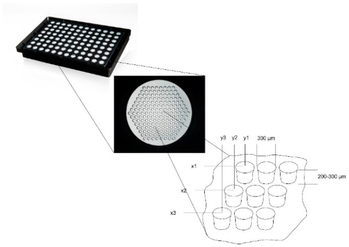
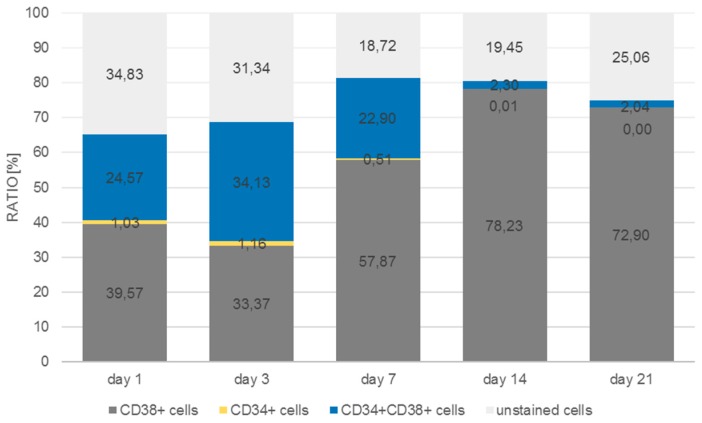
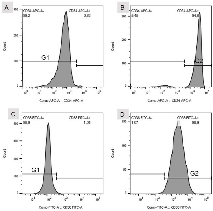
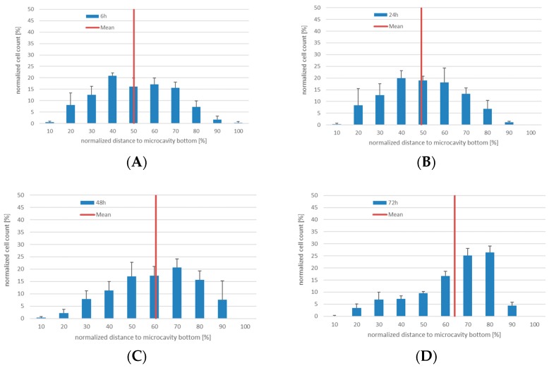
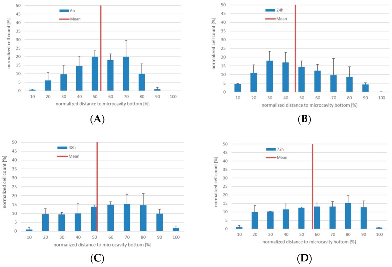
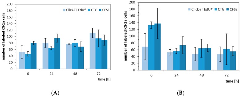
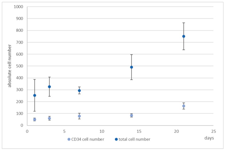
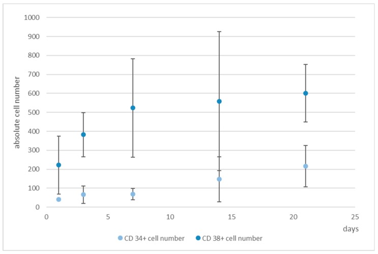
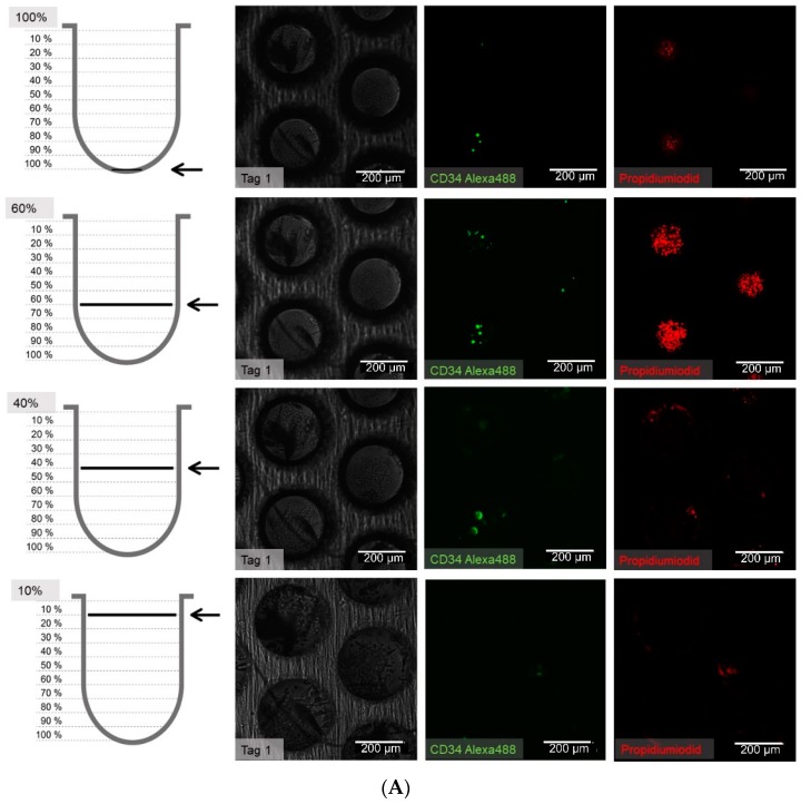
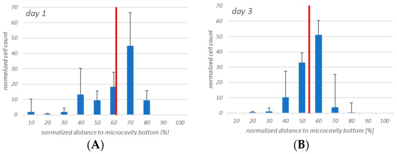
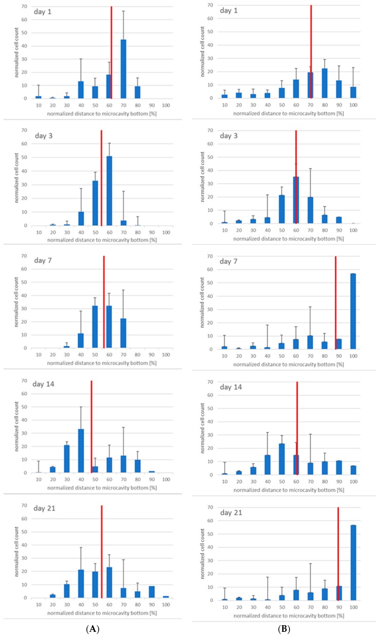
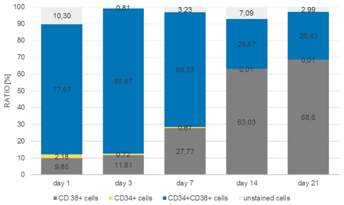
(1) Background: We describe a 4D cell culture platform with which we tried to detect and to characterize migration dynamics of single hematopoietic stem cells in polymer film microcavity arrays integrated into a microtiter plate. (2) Methods: The system was set up with CD34-expressing KG-1a cells as a surrogate for hematopoietic stem cells. We then evaluated the system as an artificial hematopoietic stem cell niche model comprised of a co-culture of human hematopoietic stem cells from cord blood (cord blood CD34 cells, hHSCs) and human mesenchymal stromal cells (hMSCs) from bone marrow over a period of 21 days. We used a software-based cell detection method to count single hematopoietic stem cells (HSCs) in microcavities. (3) Results: It was possible to detect single HSCs and their migration behavior within single microcavities. The HSCs displayed a pronounced migration behavior with one population of CD34-expressing cells located at the bottom of the microcavities and one population located in the middle of the microcavities at day 14. However, at day 21 the two populations seemed to unite again so that no clear distinction between the two was possible anymore. (4) Conclusions: Single cell migration detection was possible but microscopy and flow cytometry delivered non-uniform data sets. Further optimization is currently being developed.
(1) 背景:我们描述了一种四维细胞培养平台,利用该平台我们试图检测并表征整合到微孔板中的聚合物薄膜微腔阵列中单个造血干细胞的迁移动力学。(2) 方法:该系统以表达CD34的KG-1a细胞作为造血干细胞的替代物进行搭建。然后,我们将该系统评估为一种人工造血干细胞龛模型,该模型由来自脐带血的人类造血干细胞(脐带血CD34细胞,hHSCs)和来自骨髓的人间充质基质细胞(hMSCs)共培养21天组成。我们使用基于软件的细胞检测方法来计数微腔中的单个造血干细胞(HSCs)。(3) 结果:能够检测单个微腔内的单个HSCs及其迁移行为。在第14天,HSCs表现出明显的迁移行为,一群表达CD34的细胞位于微腔底部,另一群位于微腔中部。然而,在第21天,这两群细胞似乎又重新合并,以至于不再能清楚地区分两者。(4) 结论:单细胞迁移检测是可行的,但显微镜检查和流式细胞术提供的数据组不一致。目前正在进行进一步优化。