Faria Mun Y, Sousa David C, Almeida Bruna C, Pinto Andreia L, Ferreira Nuno P
Ophthalmology University Clinic, Faculdade de Medicina Lisboa, Universidade de Lisboa, 1649-028 Lisboa, Portugal.
Ophthalmology Department, Hospital Santa Maria, Centro Hospitalar Universitário Lisboa Norte, 1649-035 Lisboa, Portugal.
J Ophthalmol. 2019 May 2;2019:1345683. doi: 10.1155/2019/1345683. eCollection 2019.
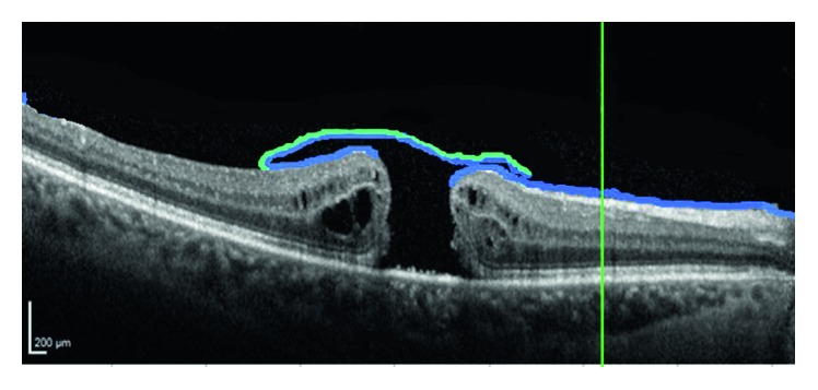
The aim of this work was to describe the ultrastructure and behavior of peeled internal limiting membrane (ILM) in macular hole (MH) surgery.
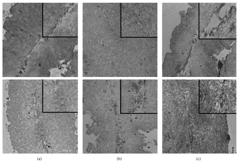
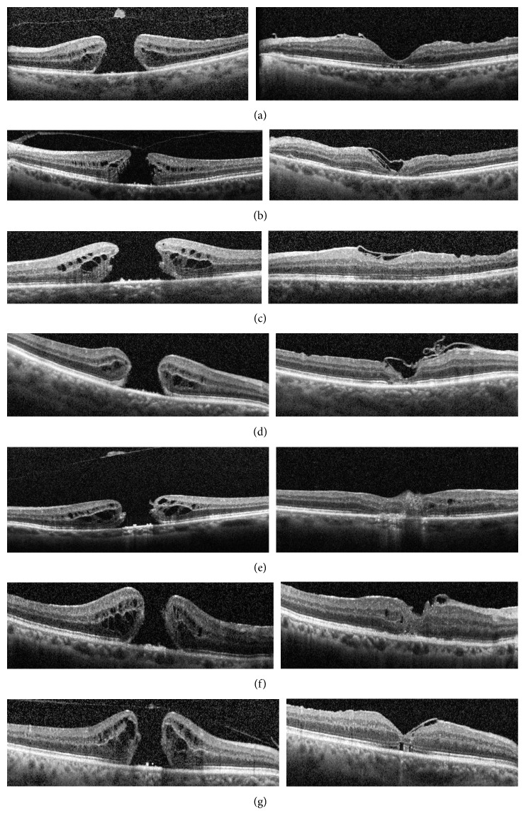
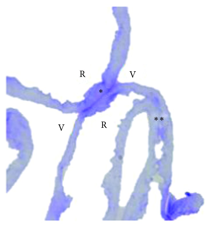
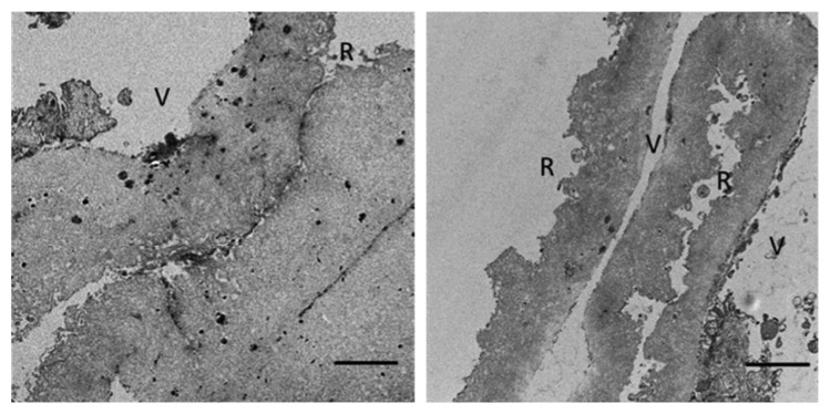
Seven patients with MH were included, and vitrectomy with ILM peeling was performed in all patients. The ILM inverted flap technique was used. Two other flaps of ILM of the same patient were collected and studied using light and transmission electron microscopy (TEM). ILM cell type, distribution, and morphology were analyzed, and the proliferation or fusion potential of the ILM interface was evaluated.
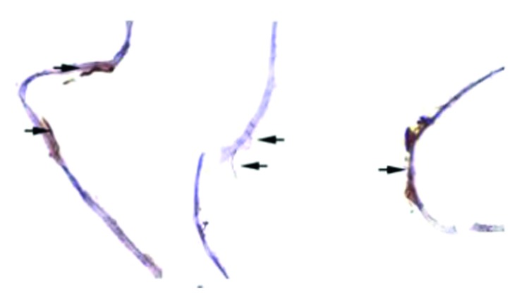
ILM vitreous sides in apposition showed signs of proliferative fibrotic activity, producing a basal membrane that merges ILM sides.
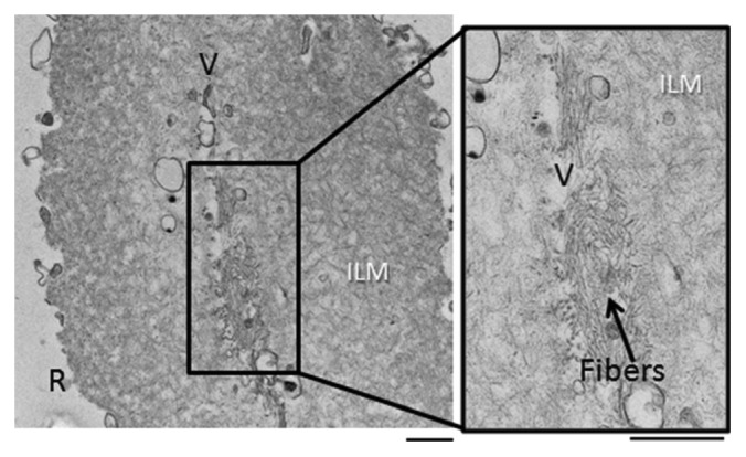
Epiretinal cells in ILM show proliferative capacity, with formation of microfibrils between adjacent sides of the ILM, which may explain adherence of ILM flaps to the hole border, contributing to closure of the hole in MH surgery. This trail is registered with NCT03799575.
本研究旨在描述黄斑裂孔(MH)手术中剥除的内界膜(ILM)的超微结构及行为。
纳入7例MH患者,所有患者均行玻璃体切除术联合ILM剥除术,采用ILM翻转瓣技术。收集同一名患者的另外两片ILM瓣,运用光学显微镜和透射电子显微镜(TEM)进行研究。分析ILM细胞类型、分布及形态,并评估ILM界面的增殖或融合潜能。
相邻的ILM玻璃体侧显示出增殖性纤维化活动迹象,产生融合ILM两侧的基底膜。
ILM中的视网膜前细胞显示出增殖能力,在ILM相邻面之间形成微原纤维,这可能解释了ILM瓣与裂孔边缘的粘连,有助于MH手术中裂孔的闭合。本试验已在ClinicalTrials.gov注册,注册号为NCT03799575。