• 文献检索
  • 文档翻译
  • 深度研究
  • 学术资讯
  • Suppr Zotero 插件Zotero 插件
  • 邀请有礼
  • 套餐&价格
  • 历史记录
应用&插件
Suppr Zotero 插件Zotero 插件浏览器插件Mac 客户端Windows 客户端微信小程序
定价
高级版会员购买积分包购买API积分包
服务
文献检索文档翻译深度研究API 文档MCP 服务
关于我们
关于 Suppr公司介绍联系我们用户协议隐私条款
关注我们

Suppr 超能文献

核心技术专利:CN118964589B侵权必究
粤ICP备2023148730 号-1Suppr @ 2026

文献检索

告别复杂PubMed语法,用中文像聊天一样搜索,搜遍4000万医学文献。AI智能推荐,让科研检索更轻松。

立即免费搜索

文件翻译

保留排版,准确专业,支持PDF/Word/PPT等文件格式,支持 12+语言互译。

免费翻译文档

深度研究

AI帮你快速写综述,25分钟生成高质量综述,智能提取关键信息,辅助科研写作。

立即免费体验

原子视角下的疟原虫肌动蛋白聚合、ATP 水解和断裂。

Atomic view into Plasmodium actin polymerization, ATP hydrolysis, and fragmentation.

机构信息

Biocenter Oulu and Faculty of Biochemistry and Molecular Medicine, University of Oulu, Oulu, Finland.

Department of Biomedicine, University of Bergen, Bergen, Norway.

出版信息

PLoS Biol. 2019 Jun 14;17(6):e3000315. doi: 10.1371/journal.pbio.3000315. eCollection 2019 Jun.

DOI:10.1371/journal.pbio.3000315
PMID:31199804
原文链接:https://pmc.ncbi.nlm.nih.gov/articles/PMC6599135/
Abstract

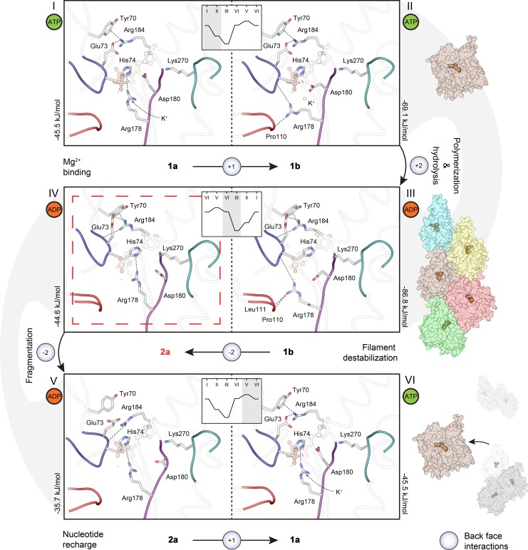
Plasmodium actins form very short filaments and have a noncanonical link between ATP hydrolysis and polymerization. Long filaments are detrimental to the parasites, but the structural factors constraining Plasmodium microfilament lengths have remained unknown. Using high-resolution crystallography, we show that magnesium binding causes a slight flattening of the Plasmodium actin I monomer, and subsequent phosphate release results in a more twisted conformation. Thus, the Mg-bound monomer is closer in conformation to filamentous (F) actin than the Ca form, and this likely facilitates polymerization. A coordinated potassium ion resides in the active site during hydrolysis and leaves together with the phosphate, a process governed by the position of the Arg178/Asp180-containing A loop. Asp180 interacts with either Lys270 or His74, depending on the protonation state of the histidine, while Arg178 links the inner and outer domains (ID and OD) of the actin protomer. Hence, the A loop acts as a switch between stable and unstable filament conformations, the latter leading to fragmentation. Our data provide a comprehensive model for polymerization, ATP hydrolysis and phosphate release, and fragmentation of parasite microfilaments. Similar mechanisms may well exist in canonical actins, although fragmentation is much less favorable due to several subtle sequence differences as well as the methylation of His73, which is absent on the corresponding His74 in Plasmodium actin I.

摘要

疟原虫肌动蛋白形成非常短的纤维,并且在 ATP 水解和聚合之间存在非典型的连接。长纤维对寄生虫有害,但限制疟原虫微丝长度的结构因素仍然未知。使用高分辨率晶体学,我们表明镁结合导致疟原虫肌动蛋白 I 单体轻微变平,随后磷酸盐释放导致更扭曲的构象。因此,与 Ca 形式相比,Mg 结合的单体在构象上更接近丝状(F)肌动蛋白,这可能促进聚合。一个协调的钾离子在水解过程中位于活性位点,并与磷酸盐一起离开,这一过程受包含 Arg178/Asp180 的 A 环的位置控制。Asp180 与 Lys270 或 His74 相互作用,具体取决于组氨酸的质子化状态,而 Arg178 将肌动蛋白原的内域(ID)和外域(OD)连接起来。因此,A 环充当稳定和不稳定纤维构象之间的开关,后者导致碎片化。我们的数据提供了一个关于聚合、ATP 水解和磷酸盐释放以及寄生虫微丝碎片化的综合模型。类似的机制可能存在于典型肌动蛋白中,尽管由于几个微妙的序列差异以及 His73 的甲基化,碎片化的可能性要小得多,而 His73 在疟原虫肌动蛋白 I 中不存在。

https://cdn.ncbi.nlm.nih.gov/pmc/blobs/3a9d/6599135/f1f2afd13a16/pbio.3000315.g007.jpg
https://cdn.ncbi.nlm.nih.gov/pmc/blobs/3a9d/6599135/acd55afc2359/pbio.3000315.g001.jpg
https://cdn.ncbi.nlm.nih.gov/pmc/blobs/3a9d/6599135/e20288a5dce9/pbio.3000315.g002.jpg
https://cdn.ncbi.nlm.nih.gov/pmc/blobs/3a9d/6599135/1f4ab82d5e41/pbio.3000315.g003.jpg
https://cdn.ncbi.nlm.nih.gov/pmc/blobs/3a9d/6599135/24b0fdb1edce/pbio.3000315.g004.jpg
https://cdn.ncbi.nlm.nih.gov/pmc/blobs/3a9d/6599135/84e91e77cf79/pbio.3000315.g005.jpg
https://cdn.ncbi.nlm.nih.gov/pmc/blobs/3a9d/6599135/c339ed2fb8ac/pbio.3000315.g006.jpg
https://cdn.ncbi.nlm.nih.gov/pmc/blobs/3a9d/6599135/f1f2afd13a16/pbio.3000315.g007.jpg

相似文献

1
Atomic view into Plasmodium actin polymerization, ATP hydrolysis, and fragmentation.原子视角下的疟原虫肌动蛋白聚合、ATP 水解和断裂。
PLoS Biol. 2019 Jun 14;17(6):e3000315. doi: 10.1371/journal.pbio.3000315. eCollection 2019 Jun.
2
Actin polymerization and ATP hydrolysis.肌动蛋白聚合与ATP水解。
Science. 1987 Oct 30;238(4827):638-44. doi: 10.1126/science.3672117.
3
Insights into the Cooperative Nature of ATP Hydrolysis in Actin Filaments.肌动蛋白丝中 ATP 水解协同性的研究进展。
Biophys J. 2018 Oct 16;115(8):1589-1602. doi: 10.1016/j.bpj.2018.08.034. Epub 2018 Sep 1.
4
A model for actin polymerization and the kinetic effects of ATP hydrolysis.肌动蛋白聚合模型及ATP水解的动力学效应。
Proc Natl Acad Sci U S A. 1985 Nov;82(21):7207-11. doi: 10.1073/pnas.82.21.7207.
5
Actin polymerization: regulation by divalent metal ion and nucleotide binding, ATP hydrolysis and binding of myosin.肌动蛋白聚合:受二价金属离子、核苷酸结合、ATP水解及肌球蛋白结合的调节。
Adv Exp Med Biol. 1994;358:71-81. doi: 10.1007/978-1-4615-2578-3_7.
6
Structural transitions of F-actin upon ATP hydrolysis at near-atomic resolution revealed by cryo-EM.冷冻电镜揭示了接近原子分辨率的 F-肌动蛋白在 ATP 水解时的结构转变。
Nat Struct Mol Biol. 2018 Jun;25(6):528-537. doi: 10.1038/s41594-018-0074-0. Epub 2018 Jun 4.
7
Structural basis for the destabilization of F-actin by phosphate release following ATP hydrolysis.ATP水解后通过磷酸释放使F-肌动蛋白不稳定的结构基础。
J Mol Biol. 1992 Oct 20;227(4):1043-53. doi: 10.1016/0022-2836(92)90520-t.
8
Actin polymerization and ATP hydrolysis.肌动蛋白聚合与ATP水解。
Adv Biophys. 1990;26:51-73. doi: 10.1016/0065-227x(90)90007-g.
9
Direct evidence for ADP-Pi-F-actin as the major intermediate in ATP-actin polymerization. Rate of dissociation of Pi from actin filaments.ADP-磷酸-肌动蛋白丝作为ATP-肌动蛋白聚合反应主要中间体的直接证据。磷酸从肌动蛋白丝上解离的速率。
Biochemistry. 1986 Dec 2;25(24):7789-92. doi: 10.1021/bi00372a001.
10
Nonlinear increase of elongation rate of actin filaments with actin monomer concentration.肌动蛋白丝伸长速率随肌动蛋白单体浓度呈非线性增加。
Biochemistry. 1986 Aug 26;25(17):4899-906. doi: 10.1021/bi00365a026.

引用本文的文献

1
Toxoplasma gondii actin filaments are tuned for rapid disassembly and turnover.刚地弓形虫肌动蛋白丝的快速解聚和周转率得到了调节。
Nat Commun. 2024 Feb 28;15(1):1840. doi: 10.1038/s41467-024-46111-3.
2
actin filaments are tuned for rapid disassembly and turnover.肌动蛋白丝经过调整以实现快速拆卸和周转。
bioRxiv. 2023 Aug 30:2023.08.29.555340. doi: 10.1101/2023.08.29.555340.
3
Sulfonylpiperazine compounds prevent Plasmodium falciparum invasion of red blood cells through interference with actin-1/profilin dynamics.磺酰基哌嗪类化合物通过干扰肌动蛋白-1/原肌球蛋白动力学来阻止恶性疟原虫入侵红细胞。

本文引用的文献

1
Mechanism of actin polymerization revealed by cryo-EM structures of actin filaments with three different bound nucleotides.三种不同结合核苷酸的肌动蛋白丝冷冻电镜结构揭示的肌动蛋白聚合机制。
Proc Natl Acad Sci U S A. 2019 Mar 5;116(10):4265-4274. doi: 10.1073/pnas.1807028115. Epub 2019 Feb 13.
2
Structural evidence for the roles of divalent cations in actin polymerization and activation of ATP hydrolysis.二价阳离子在肌动蛋白聚合和 ATP 水解激活中的作用的结构证据。
Proc Natl Acad Sci U S A. 2018 Oct 9;115(41):10345-10350. doi: 10.1073/pnas.1806394115. Epub 2018 Sep 25.
3
Inter-subunit interactions drive divergent dynamics in mammalian and Plasmodium actin filaments.
PLoS Biol. 2023 Apr 13;21(4):e3002066. doi: 10.1371/journal.pbio.3002066. eCollection 2023 Apr.
4
Structure and function of Plasmodium actin II in the parasite mosquito stages.疟原虫肌动蛋白 II 在寄生虫蚊子阶段的结构与功能。
PLoS Pathog. 2023 Mar 6;19(3):e1011174. doi: 10.1371/journal.ppat.1011174. eCollection 2023 Mar.
5
Divergent Plasmodium actin residues are essential for filament localization, mosquito salivary gland invasion and malaria transmission.不同的疟原虫肌动蛋白残基对于丝状体的定位、蚊子唾液腺入侵和疟疾传播是必不可少的。
PLoS Pathog. 2022 Aug 23;18(8):e1010779. doi: 10.1371/journal.ppat.1010779. eCollection 2022 Aug.
6
Structural basis of rapid actin dynamics in the evolutionarily divergent Leishmania parasite.进化上差异很大的利什曼原虫中肌动蛋白快速动力学的结构基础。
Nat Commun. 2022 Jun 15;13(1):3442. doi: 10.1038/s41467-022-31068-y.
7
High-resolution structures of malaria parasite actomyosin and actin filaments.疟原虫肌球蛋白和肌动蛋白丝的高分辨率结构。
PLoS Pathog. 2022 Apr 4;18(4):e1010408. doi: 10.1371/journal.ppat.1010408. eCollection 2022 Apr.
8
Structure and function of an atypical homodimeric actin capping protein from the malaria parasite.疟原虫中非典型同源二聚肌动蛋白加帽蛋白的结构与功能。
Cell Mol Life Sci. 2022 Feb 8;79(2):125. doi: 10.1007/s00018-021-04032-0.
9
The actomyosin interface contains an evolutionary conserved core and an ancillary interface involved in specificity.肌动球蛋白界面包含一个进化上保守的核心和一个参与特异性的辅助界面。
Nat Commun. 2021 Mar 25;12(1):1892. doi: 10.1038/s41467-021-22093-4.
10
Towards a structural understanding of the remodeling of the actin cytoskeleton.朝向肌动蛋白细胞骨架重构的结构理解。
Semin Cell Dev Biol. 2020 Jun;102:51-64. doi: 10.1016/j.semcdb.2019.11.018. Epub 2019 Dec 10.
亚基相互作用驱动哺乳动物和疟原虫肌动蛋白丝的不同动力学行为。
PLoS Biol. 2018 Jul 16;16(7):e2005345. doi: 10.1371/journal.pbio.2005345. eCollection 2018 Jul.
4
Structural transitions of F-actin upon ATP hydrolysis at near-atomic resolution revealed by cryo-EM.冷冻电镜揭示了接近原子分辨率的 F-肌动蛋白在 ATP 水解时的结构转变。
Nat Struct Mol Biol. 2018 Jun;25(6):528-537. doi: 10.1038/s41594-018-0074-0. Epub 2018 Jun 4.
5
Structural basis for cofilin binding and actin filament disassembly.细胞松弛素结合和肌动蛋白丝解体的结构基础。
Nat Commun. 2018 May 10;9(1):1860. doi: 10.1038/s41467-018-04290-w.
6
Motor Proteins.马达蛋白。
Cold Spring Harb Perspect Biol. 2018 May 1;10(5):a021931. doi: 10.1101/cshperspect.a021931.
7
Myosin-Driven Intracellular Transport.肌球蛋白驱动的细胞内运输。
Cold Spring Harb Perspect Biol. 2018 Mar 1;10(3):a021972. doi: 10.1101/cshperspect.a021972.
8
High-resolution cryo-EM structures of actin-bound myosin states reveal the mechanism of myosin force sensing.高分辨率冷冻电镜结构的肌球蛋白结合肌动蛋白状态揭示了肌球蛋白力感应的机制。
Proc Natl Acad Sci U S A. 2018 Feb 6;115(6):1292-1297. doi: 10.1073/pnas.1718316115. Epub 2018 Jan 22.
9
The Actin Cytoskeleton and Actin-Based Motility.肌动蛋白细胞骨架和基于肌动蛋白的运动。
Cold Spring Harb Perspect Biol. 2018 Jan 2;10(1):a018267. doi: 10.1101/cshperspect.a018267.
10
Apicomplexan actin polymerization depends on nucleation.质体锥虫肌动蛋白聚合依赖于成核。
Sci Rep. 2017 Sep 22;7(1):12137. doi: 10.1038/s41598-017-11330-w.