Mohtasib Rafat Saeed, Alshamiri Kamal Mostafa, Jobeir Aman Asad, Saidi Farida Mohsin Ambo, Masawi Ahmed Mohammed, Alabdulaziz Lamya Sami, Hussain Faisal Zaid Bin
From the Department of Biomedical Physics, Molecular and Functional Imaging, King Faisal Specialist Hospital and Research Centre, Riyadh, Saudi Arabia.
From the Department of Radilogy, King Faisal Specialist Hospital and Research Centre, Riyadh, Saudi Arabia.
Ann Saudi Med. 2019 May-Jun;39(3):143-154. doi: 10.5144/0256-4947.2019.143. Epub 2019 May 30.
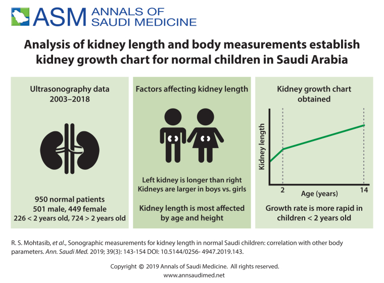
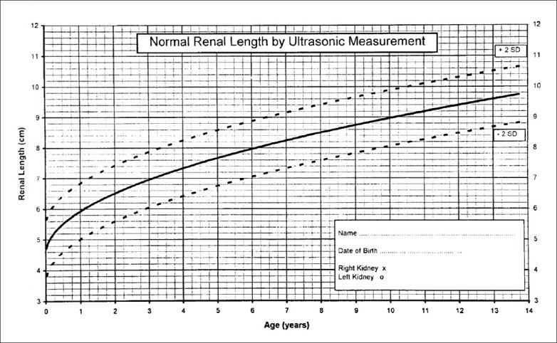
Ultrasonography provides a quick assessment of visceral organ dimensions without any risk of radiation. Since many diseases can affect the kidney size, having a reliable reference for kidney length in children is valuable for clinical assessment.
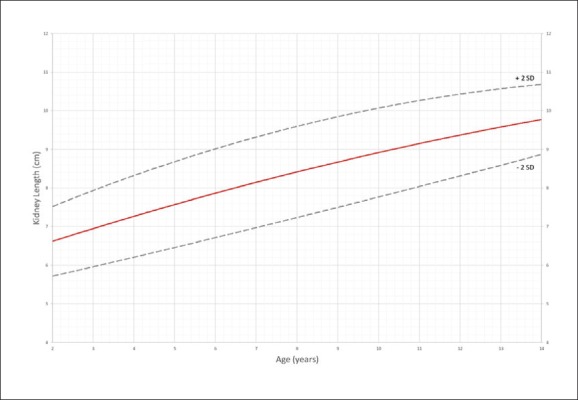
Establish normal growth curves for renal length in relation to sex, age, body weight, height, body mass index and body surface area of healthy children in Saudi Arabia.
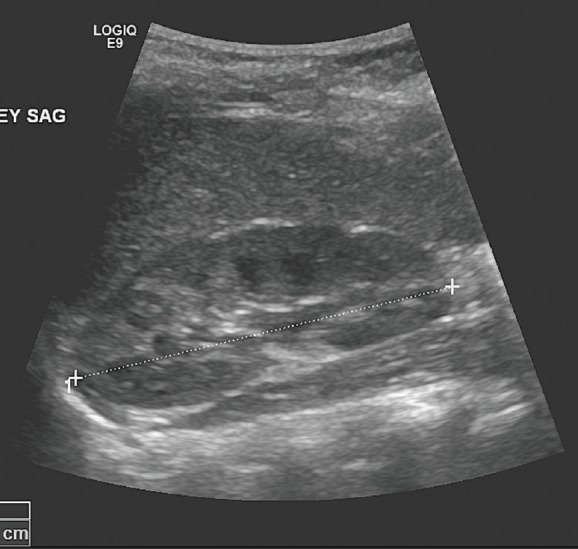
Retrospective review of ultrasonography images.
Tertiary referral hospital.
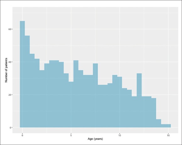
We included all normal ultrasonography exams of renal length from full-term neonates to children ≤14 years old performed between 2003 and 2018. Data was collected retrospectively from the electronic archive and patient records.
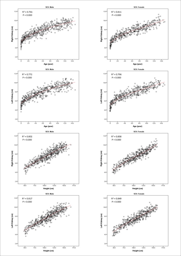
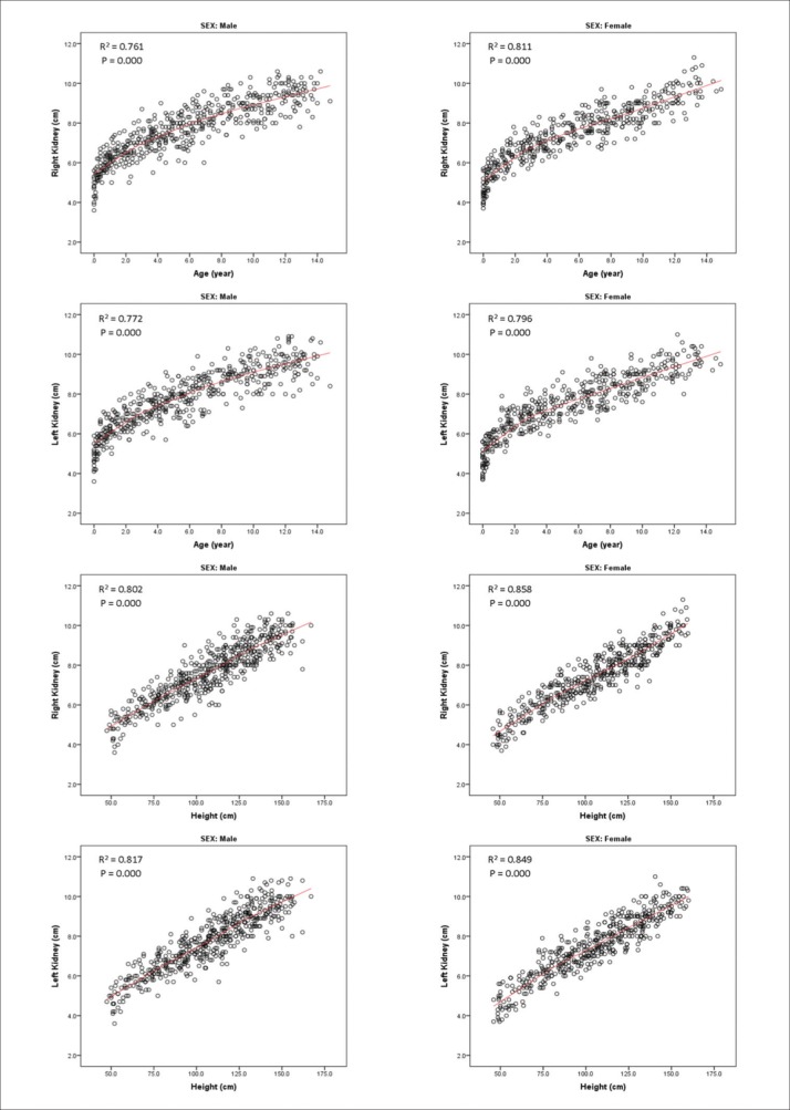
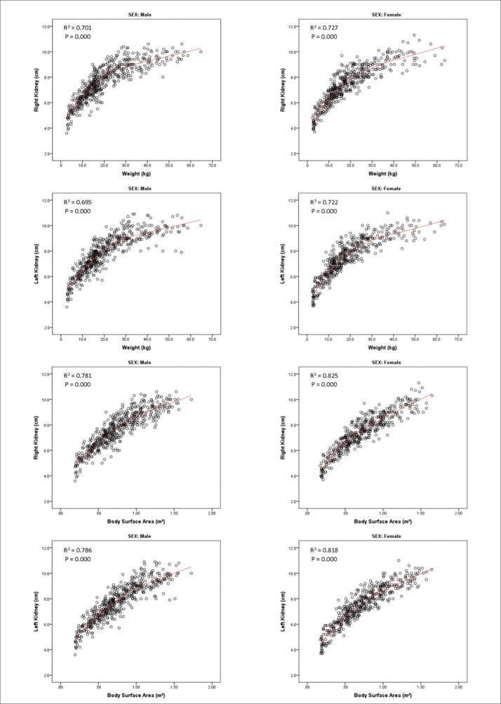
Relationship between the longitudinal length of both kidneys and age, height, weight, body mass index and body surface area.
950 patients.
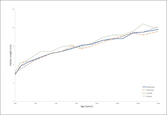
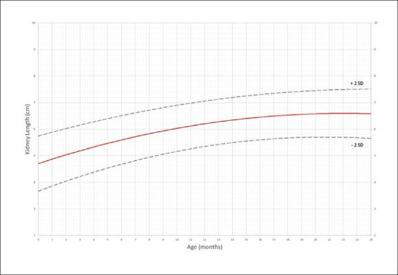
The left kidneys were longer than the right kidneys ( P<.001). Height had the most significant correlation with kidney length (R=0.829, P<.001 for right kidney; R=0.831, P<.001 for left kidney). There was a consistent difference in kidney length by sex. Both kidneys were longer in males than females ( P=.031, right kidney:, P=.015, left kidney). In terms of renal growth by age, our data showed a statistically significant difference before and after 24 months of age. There was no significant difference between populations from Saudi Arabia, Hong Kong ( P=.485) and Australia ( P=.99), but the difference between Saudi and American children was significant ( P<.001). However, we did not have the data from those studies for direct comparison. The correlation plots of renal length versus age for all four countries were similar.
The tables and correlation plots generated from this study should be useful to radiology departments in assessing conditions in children ≤14 years of age that lead to changes in renal size.
Retrospective, and there were differences in ultrasonographic techniques for patient positioning and cursor placement that can affect the reproducibility of measurements of renal length.
None.
超声检查可快速评估内脏器官大小,且无辐射风险。由于许多疾病会影响肾脏大小,因此为儿童肾脏长度建立可靠的参考标准对临床评估很有价值。
建立沙特阿拉伯健康儿童肾脏长度与性别、年龄、体重、身高、体重指数和体表面积的正常生长曲线。
对超声图像进行回顾性分析。
三级转诊医院。
我们纳入了2003年至2018年间对足月新生儿至14岁以下儿童进行的所有肾脏长度正常的超声检查。数据从电子存档和患者记录中回顾性收集。
双肾纵向长度与年龄、身高、体重、体重指数和体表面积之间的关系。
950例患者。
左肾比右肾长(P<0.001)。身高与肾脏长度的相关性最为显著(右肾R=0.829,P<0.001;左肾R=0.831,P<0.001)。肾脏长度在性别上存在一致差异。男性的双肾均比女性长(右肾P=0.031,左肾P=0.015)。就按年龄的肾脏生长情况而言,我们的数据显示24个月龄前后存在统计学显著差异。沙特阿拉伯、中国香港(P=0.485)和澳大利亚人群之间无显著差异,但沙特儿童与美国儿童之间的差异显著(P<0.001)。然而,我们没有那些研究的数据用于直接比较。所有四个国家的肾脏长度与年龄的相关性图相似。
本研究生成的表格和相关性图应有助于放射科评估14岁以下儿童导致肾脏大小变化的情况。
回顾性研究,且患者体位和光标放置的超声技术存在差异,这可能会影响肾脏长度测量的可重复性。
无。