Department of Cell and Developmental Biology, Perelman School of Medicine, University of Pennsylvania, Philadelphia, PA, United States of America.
PLoS One. 2019 Jun 20;14(6):e0218667. doi: 10.1371/journal.pone.0218667. eCollection 2019.
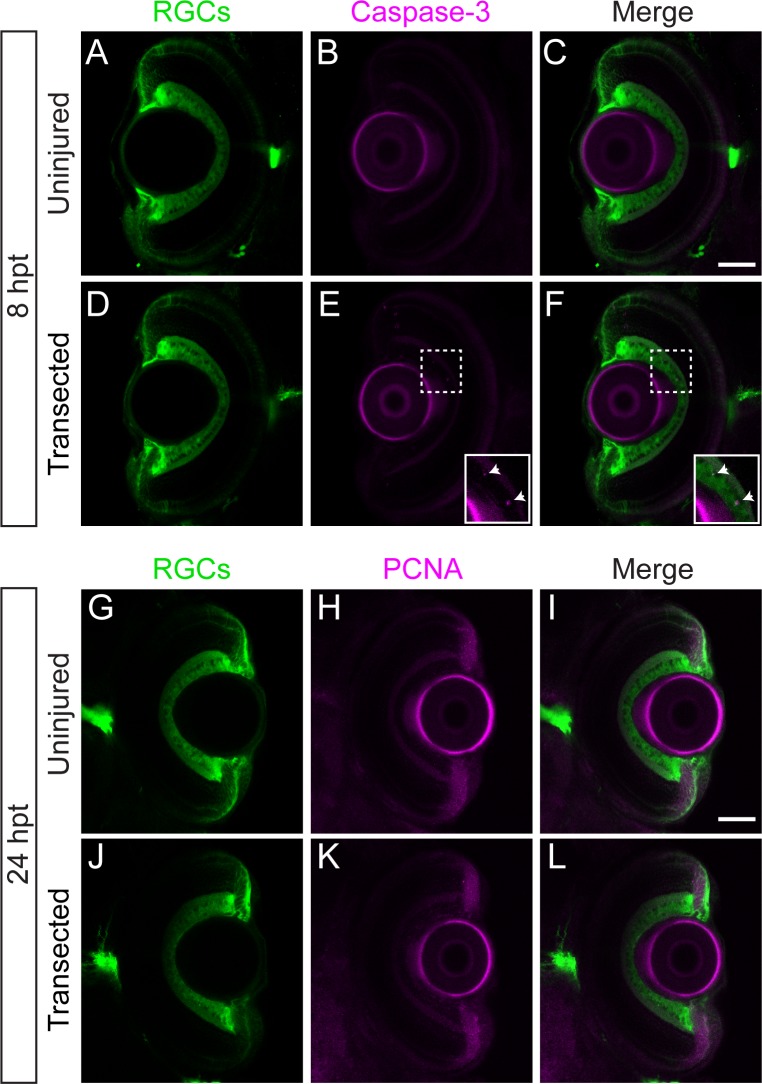
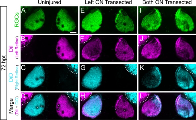
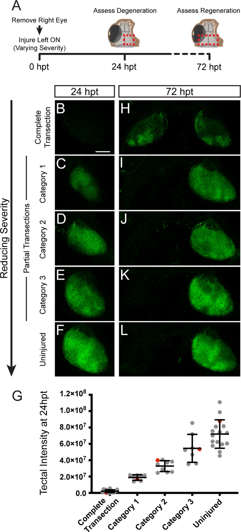
In contrast to mammals, retinal ganglion cells (RGC) axons of the optic nerve even in mature zebrafish exhibit a remarkable capacity for spontaneous regeneration. One constraint of using adult zebrafish is the limited ability to visualize the regeneration process in live animals. To dynamically visualize and trace the degree of target specific optic nerve regeneration, we took advantage of the optical transparency still preserved in post developmental larval zebrafish. We developed a rapid and robust assay to physically transect the larval optic nerve and find that by 96 hours post injury RGC axons have robustly regrown onto the optic tectum. We observe functional regeneration by 8 days post injury, and demonstrate that similar to adult zebrafish, optic nerve transection in larval zebrafish does not prominently induce cell death or proliferation of RGC neurons. Furthermore, we find that partial optic nerve transection results in axonal growth predominantly to the original, contralateral tectum, while complete transection results in innervation of both the correct contralateral and 'incorrect' ipsilateral tectum. Axonal tracing reveals that although regenerating axons innervate the 'incorrect' ipsilateral tectum, they successfully target their topographically appropriate synaptic areas. Combined, our results validate post developmental larval zebrafish as a powerful model for optic nerve regeneration, and reveal intricate mechanistic differences between axonal growth, midline guidance and synaptic targeting during zebrafish optic nerve regeneration.
与哺乳动物不同,即使在成熟的斑马鱼中,视神经中的视网膜神经节细胞 (RGC) 轴突也表现出惊人的自发再生能力。使用成年斑马鱼的一个限制是,在活体动物中观察再生过程的能力有限。为了动态可视化和追踪特定目标视神经再生的程度,我们利用了仍在发育后幼虫斑马鱼中保留的光学透明度。我们开发了一种快速而稳健的测定法来物理横切幼虫视神经,并发现 RGC 轴突在损伤后 96 小时已经强劲地再生到视顶盖。我们在损伤后 8 天观察到功能再生,并证明与成年斑马鱼相似,幼虫斑马鱼视神经横切不会明显诱导 RGC 神经元死亡或增殖。此外,我们发现部分视神经横切导致轴突生长主要到原始的对侧视顶盖,而完全横切导致正确的对侧和“错误”的同侧视顶盖的神经支配。轴突追踪表明,尽管再生轴突支配“错误”的同侧视顶盖,但它们成功地将其投射到适当的拓扑突触区域。总之,我们的结果验证了发育后幼虫斑马鱼是视神经再生的有力模型,并揭示了斑马鱼视神经再生过程中轴突生长、中线引导和突触靶向之间的复杂机制差异。