Marquart Gregory D, Tabor Kathryn M, Horstick Eric J, Brown Mary, Geoca Alexandra K, Polys Nicholas F, Nogare Damian Dalle, Burgess Harold A
Division of Developmental Biology, Eunice Kennedy Shriver National Institute of Child Health and Human Development, Building 6B, Room: 3B-308, 6 Center Dr., Bethesda, MD 20892-0002.
Neuroscience and Cognitive Science Program, University of Maryland, College Park, MD 20742.
Gigascience. 2017 Aug 1;6(8):1-15. doi: 10.1093/gigascience/gix056.
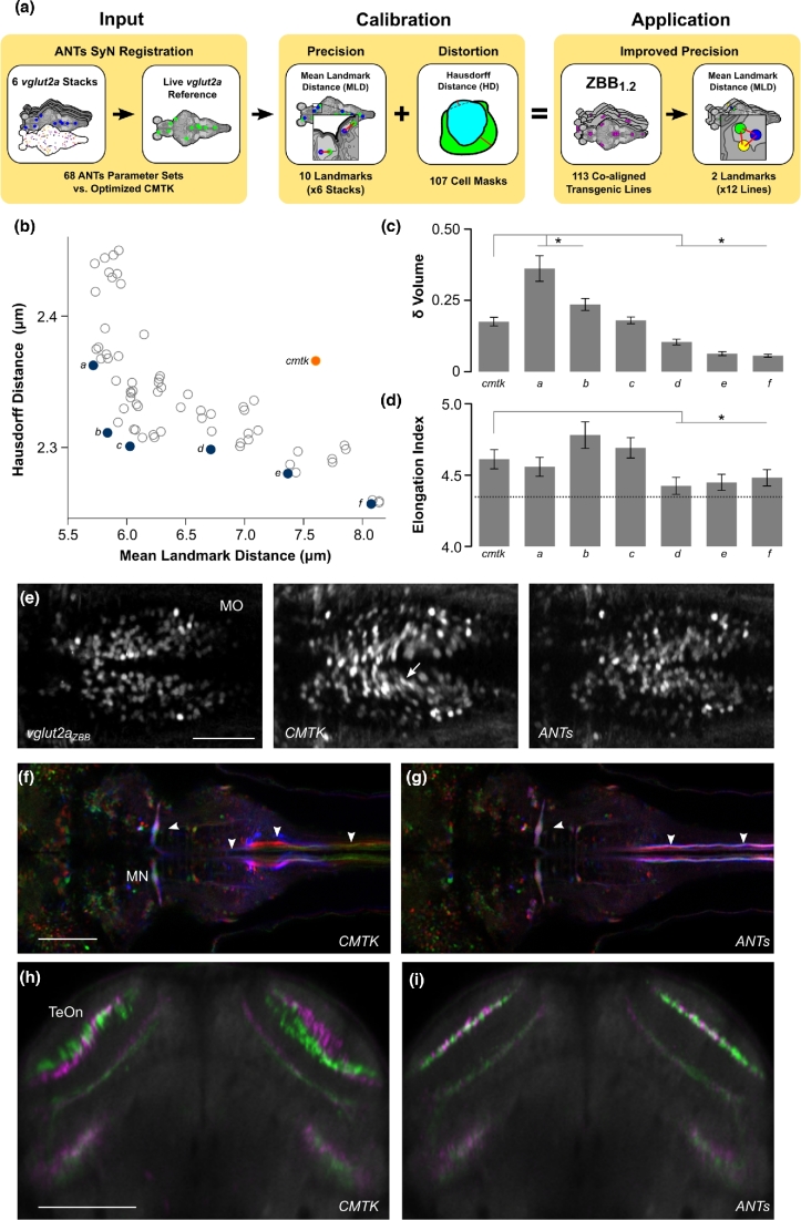
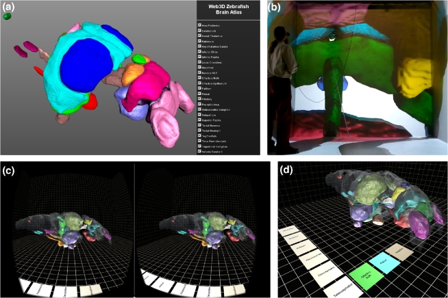
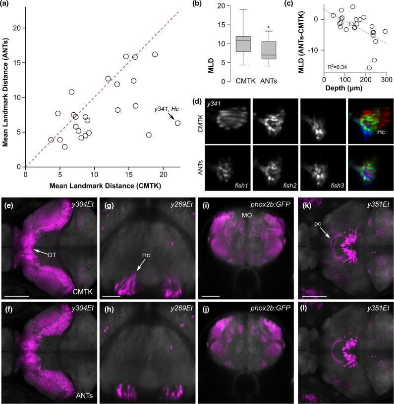
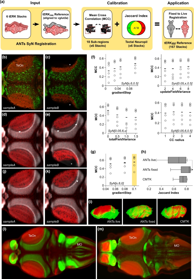
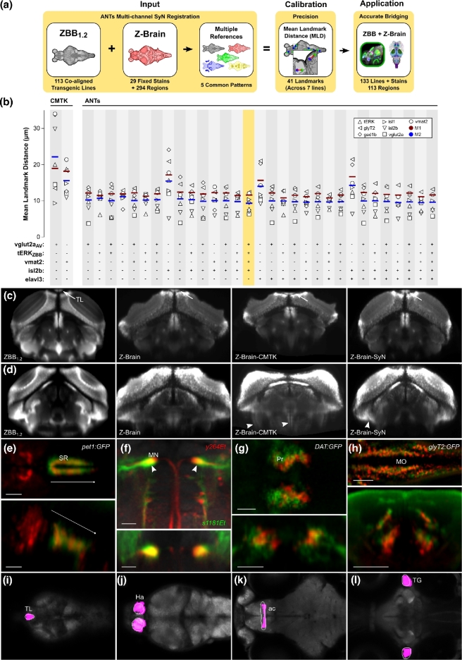
Atlases provide a framework for spatially mapping information from diverse sources into a common reference space. Specifically, brain atlases allow annotation of gene expression, cell morphology, connectivity, and activity. In larval zebrafish, advances in genetics, imaging, and computational methods now allow the collection of such information brain-wide. However, due to technical considerations, disparate datasets may use different references and may not be aligned to the same coordinate space. Two recent larval zebrafish atlases exemplify this problem: Z-Brain, containing gene expression, neural activity, and neuroanatomical segmentations, was acquired using immunohistochemical stains, while the Zebrafish Brain Browser (ZBB) was constructed from live scans of fluorescent reporters in transgenic larvae. Although different references were used, the atlases included several common transgenic patterns that provide potential "bridges" for transforming each into the other's coordinate space. We tested multiple bridging channels and registration algorithms and found that the symmetric diffeomorphic normalization algorithm improved live brain registration precision while better preserving cell morphology than B-spline-based registrations. Symmetric diffeomorphic normalization also corrected for tissue distortion introduced during fixation. Multi-reference channel optimization provided a transformation that enabled Z-Brain and ZBB to be co-aligned with precision of approximately a single cell diameter and minimal perturbation of cell and tissue morphology. Finally, we developed software to visualize brain regions in 3 dimensions, including a virtual reality neuroanatomy explorer. This study demonstrates the feasibility of integrating whole brain datasets, despite disparate reference templates and acquisition protocols, when sufficient information is present for bridging. Increased accuracy and interoperability of zebrafish digital brain atlases will facilitate neurobiological studies.
图谱为将来自不同来源的信息在空间上映射到一个共同的参考空间提供了框架。具体而言,脑图谱允许对基因表达、细胞形态、连接性和活性进行注释。在斑马鱼幼体中,遗传学、成像和计算方法的进步现在使得能够在全脑范围内收集此类信息。然而,由于技术方面的考虑,不同的数据集可能使用不同的参考,并且可能未对齐到相同的坐标空间。最近的两个斑马鱼幼体图谱就体现了这个问题:包含基因表达、神经活动和神经解剖学分割的Z - Brain是使用免疫组织化学染色获得的,而斑马鱼脑浏览器(ZBB)是由转基因幼体中荧光报告基因的实时扫描构建而成。尽管使用了不同的参考,但这些图谱包含了几种常见的转基因模式,为将彼此转换到对方的坐标空间提供了潜在的“桥梁”。我们测试了多种桥接通道和配准算法,发现对称微分同胚归一化算法提高了活体脑配准精度,同时比基于B样条的配准更好地保留了细胞形态。对称微分同胚归一化还校正了固定过程中引入的组织变形。多参考通道优化提供了一种变换,使Z - Brain和ZBB能够以大约单个细胞直径的精度共对齐,并且对细胞和组织形态的扰动最小。最后,我们开发了用于三维可视化脑区的软件,包括一个虚拟现实神经解剖学浏览器。这项研究证明了,当存在足够的信息用于桥接时,尽管参考模板和采集协议不同,整合全脑数据集是可行的。斑马鱼数字脑图谱准确性和互操作性的提高将促进神经生物学研究。