• 文献检索
  • 文档翻译
  • 深度研究
  • 学术资讯
  • Suppr Zotero 插件Zotero 插件
  • 邀请有礼
  • 套餐&价格
  • 历史记录
应用&插件
Suppr Zotero 插件Zotero 插件浏览器插件Mac 客户端Windows 客户端微信小程序
定价
高级版会员购买积分包购买API积分包
服务
文献检索文档翻译深度研究API 文档MCP 服务
关于我们
关于 Suppr公司介绍联系我们用户协议隐私条款
关注我们

Suppr 超能文献

核心技术专利:CN118964589B侵权必究
粤ICP备2023148730 号-1Suppr @ 2026

文献检索

告别复杂PubMed语法,用中文像聊天一样搜索,搜遍4000万医学文献。AI智能推荐,让科研检索更轻松。

立即免费搜索

文件翻译

保留排版,准确专业,支持PDF/Word/PPT等文件格式,支持 12+语言互译。

免费翻译文档

深度研究

AI帮你快速写综述,25分钟生成高质量综述,智能提取关键信息,辅助科研写作。

立即免费体验

用中性红染料追踪小鼠中枢神经系统髓鞘再生病变。

Tracking the evolution of CNS remyelinating lesion in mice with neutral red dye.

机构信息

Department of Biology, Georgetown University, Washington, DC 20057.

Department of Biology, Georgetown University, Washington, DC 20057;

出版信息

Proc Natl Acad Sci U S A. 2019 Jul 9;116(28):14290-14299. doi: 10.1073/pnas.1819343116. Epub 2019 Jun 24.

DOI:10.1073/pnas.1819343116
PMID:31235582
原文链接:https://pmc.ncbi.nlm.nih.gov/articles/PMC6628798/
Abstract

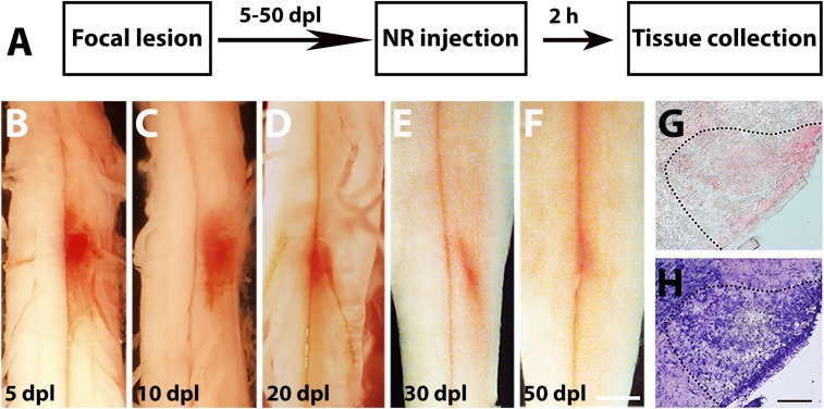
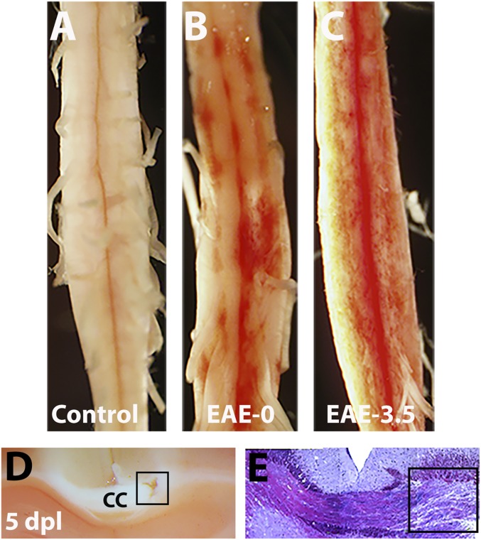
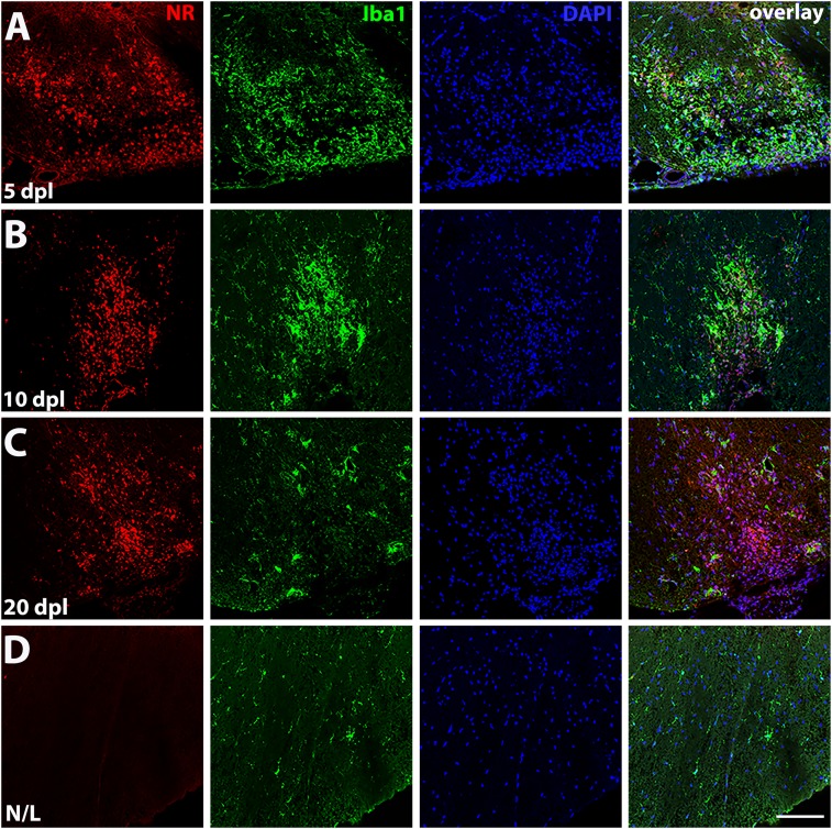
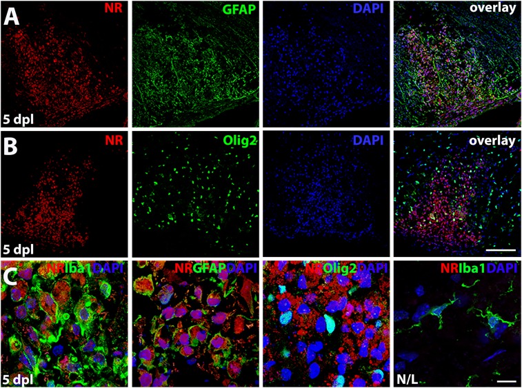
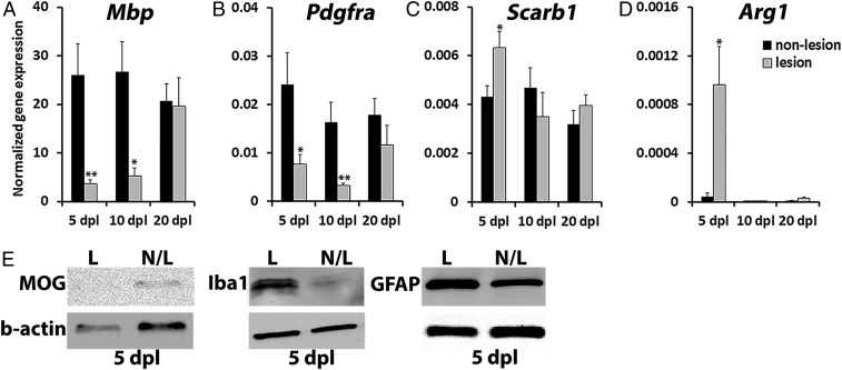
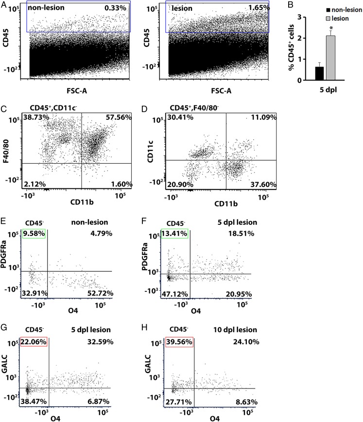
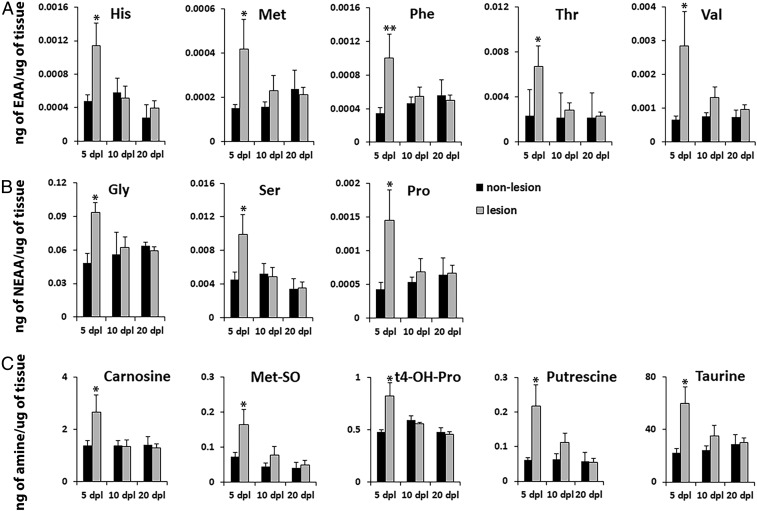
Animal models of central nervous system (CNS) demyelination, including toxin-induced focal demyelination and immune-mediated demyelination through experimental autoimmune encephalomyelitis (EAE), have provided valuable insights into the mechanisms of neuroinflammation and CNS remyelination. However, the ability to track changes in transcripts, proteins, and metabolites, as well as cellular populations during the evolution of a focal lesion, has remained challenging. Here, we developed a method to label CNS demyelinating lesions by the intraperitoneal injection of a vital dye, neutral red (NR), into mice before killing. We demonstrate that NR-labeled lesions can be easily identified on the intact spinal cord in both lysolecithin- and EAE-mediated demyelination models. Using fluorescence microscopy, we detected NR in activated macrophages/microglia and astrocytes, but not in oligodendrocytes present in lesions. Importantly, we successfully performed RT-qPCR, Western blot, flow cytometry, and mass spectrometry analysis of precisely dissected NR-labeled lesions at 5, 10, and 20 d postlesion (dpl) and found differential changes in transcripts, proteins, cell populations, and metabolites in lesions over the course of remyelination. Therefore, NR administration is a simple and powerful method to track and analyze the detailed molecular, cellular, and metabolic changes that occur within the lesion microenvironment over time following CNS injury. Furthermore, this method can be used to identify molecular and metabolic pathways that regulate neuroinflammation and remyelination and facilitate the development of therapies to promote repair in demyelinating disorders such as multiple sclerosis.

摘要

中枢神经系统(CNS)脱髓鞘的动物模型,包括毒素诱导的局灶性脱髓鞘和通过实验性自身免疫性脑脊髓炎(EAE)诱导的免疫介导的脱髓鞘,为神经炎症和 CNS 髓鞘再生的机制提供了有价值的见解。然而,在局灶性病变的发展过程中,跟踪转录物、蛋白质和代谢物以及细胞群体变化的能力仍然具有挑战性。在这里,我们开发了一种在杀死小鼠之前通过腹腔内注射活染料中性红(NR)来标记 CNS 脱髓鞘病变的方法。我们证明,NR 标记的病变可以在溶磷脂酶和 EAE 介导的脱髓鞘模型中的完整脊髓上轻松识别。使用荧光显微镜,我们在激活的巨噬细胞/小胶质细胞和星形胶质细胞中检测到 NR,但在病变中存在的少突胶质细胞中未检测到 NR。重要的是,我们成功地对 5、10 和 20 天病变后(dpl)精确解剖的 NR 标记病变进行了 RT-qPCR、Western blot、流式细胞术和质谱分析,发现髓鞘再生过程中病变中转录物、蛋白质、细胞群体和代谢物发生了差异变化。因此,NR 给药是一种简单而强大的方法,可以跟踪和分析 CNS 损伤后随时间推移在病变微环境中发生的详细分子、细胞和代谢变化。此外,该方法可用于鉴定调节神经炎症和髓鞘再生的分子和代谢途径,并促进促进脱髓鞘疾病(如多发性硬化症)修复的治疗方法的开发。

https://cdn.ncbi.nlm.nih.gov/pmc/blobs/09e4/6628798/45cca7cd1939/pnas.1819343116fig07.jpg
https://cdn.ncbi.nlm.nih.gov/pmc/blobs/09e4/6628798/18fa08e76b0b/pnas.1819343116fig01.jpg
https://cdn.ncbi.nlm.nih.gov/pmc/blobs/09e4/6628798/f39dcc8f69d1/pnas.1819343116fig02.jpg
https://cdn.ncbi.nlm.nih.gov/pmc/blobs/09e4/6628798/79ed9665e806/pnas.1819343116fig03.jpg
https://cdn.ncbi.nlm.nih.gov/pmc/blobs/09e4/6628798/d64aa6bd362e/pnas.1819343116fig04.jpg
https://cdn.ncbi.nlm.nih.gov/pmc/blobs/09e4/6628798/cf82e58b9bfe/pnas.1819343116fig05.jpg
https://cdn.ncbi.nlm.nih.gov/pmc/blobs/09e4/6628798/6bc764d442b0/pnas.1819343116fig06.jpg
https://cdn.ncbi.nlm.nih.gov/pmc/blobs/09e4/6628798/45cca7cd1939/pnas.1819343116fig07.jpg

相似文献

1
Tracking the evolution of CNS remyelinating lesion in mice with neutral red dye.用中性红染料追踪小鼠中枢神经系统髓鞘再生病变。
Proc Natl Acad Sci U S A. 2019 Jul 9;116(28):14290-14299. doi: 10.1073/pnas.1819343116. Epub 2019 Jun 24.
2
Failure of remyelination in the nonhuman primate optic nerve.非人灵长类动物视神经中髓鞘再生失败。
Brain Pathol. 2005 Jul;15(3):198-207. doi: 10.1111/j.1750-3639.2005.tb00521.x.
3
Acutely damaged axons are remyelinated in multiple sclerosis and experimental models of demyelination.在多发性硬化症和脱髓鞘实验模型中,急性受损的轴突会重新髓鞘化。
Glia. 2017 Aug;65(8):1350-1360. doi: 10.1002/glia.23167. Epub 2017 May 31.
4
Fibronectin aggregation in multiple sclerosis lesions impairs remyelination.纤维连接蛋白在多发性硬化病变中的聚集会损害髓鞘的修复。
Brain. 2013 Jan;136(Pt 1):116-31. doi: 10.1093/brain/aws313.
5
CNS-resident glial progenitor/stem cells produce Schwann cells as well as oligodendrocytes during repair of CNS demyelination.中枢神经系统驻留的神经胶质前体细胞/干细胞在中枢神经系统脱髓鞘修复过程中产生施万细胞和少突胶质细胞。
Cell Stem Cell. 2010 Jun 4;6(6):578-90. doi: 10.1016/j.stem.2010.04.002.
6
miR-219 Cooperates with miR-338 in Myelination and Promotes Myelin Repair in the CNS.miR-219在髓鞘形成过程中与miR-338协同作用,并促进中枢神经系统中的髓鞘修复。
Dev Cell. 2017 Mar 27;40(6):566-582.e5. doi: 10.1016/j.devcel.2017.03.001.
7
The role of oligodendrocytes and oligodendrocyte progenitors in CNS remyelination.少突胶质细胞和少突胶质前体细胞在中枢神经系统髓鞘再生中的作用。
Adv Exp Med Biol. 1999;468:183-97. doi: 10.1007/978-1-4615-4685-6_15.
8
Drug-based modulation of endogenous stem cells promotes functional remyelination in vivo.基于药物的内源性干细胞调节促进体内功能性髓鞘再生。
Nature. 2015 Jun 11;522(7555):216-20. doi: 10.1038/nature14335. Epub 2015 Apr 20.
9
Leonurine suppresses neuroinflammation through promoting oligodendrocyte maturation.益母草碱通过促进少突胶质细胞成熟来抑制神经炎症。
J Cell Mol Med. 2019 Feb;23(2):1470-1485. doi: 10.1111/jcmm.14053. Epub 2018 Dec 16.
10
Experimental demyelination and remyelination of murine spinal cord by focal injection of lysolecithin.通过局部注射溶血卵磷脂对小鼠脊髓进行实验性脱髓鞘和再髓鞘化。
J Vis Exp. 2015 Mar 26(97):52679. doi: 10.3791/52679.

引用本文的文献

1
PAK1 regulates oligodendroglial proliferation and repopulation in homeostatic and demyelinating brain.PAK1在稳态和脱髓鞘脑内调节少突胶质细胞的增殖和再填充。
Cell Mol Life Sci. 2025 Jun 28;82(1):260. doi: 10.1007/s00018-025-05728-3.
2
Type I collagen secreted in white matter lesions inhibits remyelination and functional recovery.在白质病变中分泌的I型胶原蛋白会抑制髓鞘再生和功能恢复。
Cell Death Dis. 2025 Apr 13;16(1):285. doi: 10.1038/s41419-025-07633-w.
3
Intrinsic retinoic acid synthesis is required for oligodendrocyte progenitor expansion during CNS remyelination.

本文引用的文献

1
Carnosine improves functional recovery and structural regeneration after sciatic nerve crush injury in rats.肌肽可改善大鼠坐骨神经挤压伤后的功能恢复和结构再生。
Life Sci. 2018 Dec 15;215:22-30. doi: 10.1016/j.lfs.2018.10.043. Epub 2018 Nov 1.
2
Carnosine as an effective neuroprotector in brain pathology and potential neuromodulator in normal conditions.肌肽作为一种有效的神经保护剂在脑部病理学中的应用,以及在正常条件下作为一种潜在的神经调节剂。
Amino Acids. 2019 Jan;51(1):139-150. doi: 10.1007/s00726-018-2667-7. Epub 2018 Oct 23.
3
Therapeutic potential of carbonyl-scavenging carnosine derivative in metabolic disorders.
在中枢神经系统再髓鞘化过程中,少突胶质细胞祖细胞的扩增需要内源性视黄酸的合成。
Front Cell Neurosci. 2025 Feb 24;19:1550139. doi: 10.3389/fncel.2025.1550139. eCollection 2025.
4
Senescent-like microglia limit remyelination through the senescence associated secretory phenotype.衰老样小胶质细胞通过衰老相关分泌表型限制髓鞘再生。
Nat Commun. 2025 Mar 7;16(1):2283. doi: 10.1038/s41467-025-57632-w.
5
Forceps minor control of social behaviour.小镊子对社会行为的控制。 (不过感觉这个表述不太符合正常医学语境逻辑,你可以检查下原文是否准确)
Sci Rep. 2024 Dec 16;14(1):30492. doi: 10.1038/s41598-024-81930-w.
6
Fueling neurodegeneration: metabolic insights into microglia functions.为神经退行性疾病提供燃料:对小胶质细胞功能的代谢见解。
J Neuroinflammation. 2024 Nov 17;21(1):300. doi: 10.1186/s12974-024-03296-0.
7
Neutral Red Labeling: A Novel Vital Staining Method for Investigating Central and Peripheral Nervous System Lesions.中性红染色:一种用于研究中枢和外周神经系统损伤的新型活体染色方法。
Acta Histochem Cytochem. 2024 Aug 29;57(4):131-135. doi: 10.1267/ahc.24-00038. Epub 2024 Aug 23.
8
Spatial dynamics of mammalian brain development and neuroinflammation by multimodal tri-omics mapping.通过多模态三基因组图谱绘制研究哺乳动物大脑发育和神经炎症的空间动态
bioRxiv. 2024 Jul 28:2024.07.28.605493. doi: 10.1101/2024.07.28.605493.
9
Control of OPC proliferation and repopulation by the intellectual disability gene PAK1 under homeostatic and demyelinating conditions.在稳态和脱髓鞘条件下,智力残疾基因PAK1对少突胶质前体细胞增殖和再填充的调控
bioRxiv. 2024 Apr 29:2024.04.26.591153. doi: 10.1101/2024.04.26.591153.
10
The Mouse Model of Internal Capsule Demyelination: A Novel Tool for Investigating Motor Functional Changes Caused by Demyelination and for Evaluating Drugs That Promote Remyelination.内囊脱髓鞘小鼠模型:一种用于研究脱髓鞘引起的运动功能变化以及评估促进髓鞘再生药物的新型工具。
Acta Histochem Cytochem. 2024 Feb 29;57(1):1-5. doi: 10.1267/ahc.24-00005. Epub 2024 Feb 23.
羰基清除型肌肽衍生物在代谢紊乱中的治疗潜力。
J Clin Invest. 2018 Dec 3;128(12):5198-5200. doi: 10.1172/JCI124304. Epub 2018 Oct 22.
4
Oligodendrocyte Bioenergetics in Health and Disease.少突胶质细胞的生物能量学:在健康与疾病中的作用
Neuroscientist. 2019 Aug;25(4):334-343. doi: 10.1177/1073858418793077. Epub 2018 Aug 20.
5
Multiple Sclerosis.多发性硬化症
N Engl J Med. 2018 Jan 11;378(2):169-180. doi: 10.1056/NEJMra1401483.
6
Metabolomics-based discovery of a metabolite that enhances oligodendrocyte maturation.基于代谢组学发现一种促进少突胶质细胞成熟的代谢物。
Nat Chem Biol. 2018 Jan;14(1):22-28. doi: 10.1038/nchembio.2517. Epub 2017 Nov 13.
7
Unconventional Myosin ID is Involved in Remyelination After Cuprizone-Induced Demyelination.非典型肌球蛋白 ID 参与了 cuprizone 诱导脱髓鞘后的髓鞘再生。
Neurochem Res. 2018 Jan;43(1):195-204. doi: 10.1007/s11064-017-2413-7. Epub 2017 Oct 6.
8
Remyelination therapies: a new direction and challenge in multiple sclerosis.髓鞘修复治疗:多发性硬化症的新方向和挑战。
Nat Rev Drug Discov. 2017 Sep;16(9):617-634. doi: 10.1038/nrd.2017.115. Epub 2017 Jul 7.
9
Creatine Enhances Mitochondrial-Mediated Oligodendrocyte Survival After Demyelinating Injury.肌酸可增强脱髓鞘损伤后线粒体介导的少突胶质细胞存活能力。
J Neurosci. 2017 Feb 8;37(6):1479-1492. doi: 10.1523/JNEUROSCI.1941-16.2016. Epub 2017 Jan 9.
10
IL4I1 augments CNS remyelination and axonal protection by modulating T cell driven inflammation.白细胞介素4诱导分子1(IL4I1)通过调节T细胞驱动的炎症反应增强中枢神经系统的髓鞘再生和轴突保护。
Brain. 2016 Dec;139(Pt 12):3121-3136. doi: 10.1093/brain/aww254. Epub 2016 Oct 25.