Department of Cardiology, "Victor Babes" University of Medicine and Pharmacy, 300041 Timisoara, Romania.
Department of Functional Sciences, "Victor Babes" University of Medicine and Pharmacy, 300041 Timisoara, Romania.
Medicina (Kaunas). 2019 Jun 25;55(6):309. doi: 10.3390/medicina55060309.
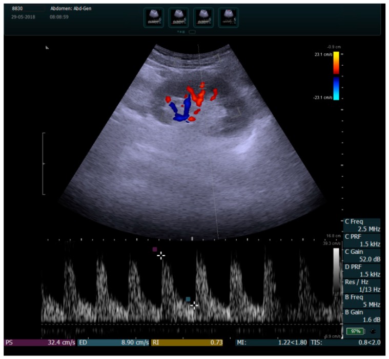
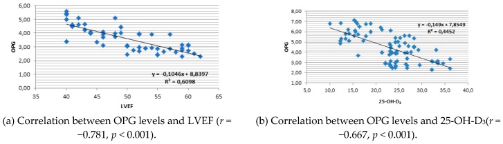
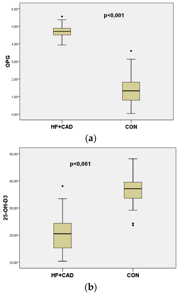
: The purpose of the study is to correlate vascular calcification biomarkers osteoprotegerin (OPG) and 25-hydroxyvitamin D (25-OH-D), indicators of arterial stiffness carotid-femoral pulse wave velocity (c-f PWV) and renal resistive index (RRI), with parameters of left ventricular function in heart failure patients versus control. : Our case-control study compared 60 patients with ischemic heart failure and reduced left ventricular ejection fraction (LVEF) (<40%) with a control group of 60 healthy age-matched subjects (CON). Serum levels of OPG and 25-OH-D were determined by ELISA. Left ventricular volumes (LVESV, LVEDV) and LVEF were measured by echocardiography. C-f PWV was determined using the arteriograph device. RRI was measured by duplex Doppler. Peak systolic velocity (PSV) and minimum end-diastolic velocity (EDV) were determined using angle correction. The estimated glomerular filtration rate (eGFR) was calculated using the MDRD equation. The Pearson's correlation coefficient was used for interpretation of results. : OPG values were significantly higher in heart failure (HF) patients vs. CON (4.7 ± 0.25 vs. 1.3 ± 0.67 ng/mL, < 0.001). 25-OH vitamin D levels were significantly lower in HF patients vs. CON (20.49 ± 7.31 vs. 37.09 ± 4.59 ng/mL, < 0.001). Multiple regression analysis considering 25-OH D3 as a dependent variable demonstrated indicators of vascular stiffness RRI, c-f PWV and vascular calcification biomarker OPG as predictors. OPG values were significantly correlated with cardiac parameters LVEDV ( = 0.862, < 0.001), LVEF ( = -0.832, < 0.001), and c-f PWV( = 0.833, < 0.001), and also with 25-OH-D ( = -0.636, < 0.001). RRI values were significantly correlated with cardiac parameters LVEDV ( = 0.586, < 0.001) and LVEF ( = -0.587, < 0.001), and with eGFR ( = -0.488, < 0.001), c-f PWV( = 0.640, < 0.001), and 25-OH-D ( = -0.732, < 0.001). : This study showed significant correlations between vitamin D deficit and vascular stiffness indicators in heart failure patients with reduced ejection fraction, demonstrating the importance of these examinations for a better evaluation of these patients. Together with the evaluation of renal function, the measurement of vascular stiffness indicators and biomarkers might play a key role in identifying patients at greater risk for worsening disease prognosis and for shorter life expectancy, who could benefit from vitamin D supplementation. The abstract was accepted for presentation at the Congress of the European Society of Cardiology, Munich, 2018.
: 研究目的是将血管钙化标志物骨保护素(OPG)和 25-羟维生素 D(25-OH-D)与颈动脉-股动脉脉搏波速度(c-f PWV)和肾血管阻力指数(RRI)等动脉僵硬度指标与心力衰竭患者的左心室功能参数相关联。:我们的病例对照研究比较了 60 例缺血性心力衰竭和左心室射血分数(LVEF)降低(<40%)的患者与 60 例年龄匹配的健康对照组(CON)。通过 ELISA 测定血清 OPG 和 25-OH-D 水平。通过超声心动图测量左心室容量(LVESV、LVEDV)和 LVEF。使用动脉造影仪测定 c-f PWV。使用双功能多普勒测定 RRI。通过角度校正确定收缩期峰值速度(PSV)和最小舒张末期速度(EDV)。使用 MDRD 方程计算估计肾小球滤过率(eGFR)。使用 Pearson 相关系数解释结果。:心力衰竭(HF)患者的 OPG 值明显高于 CON(4.7±0.25 与 1.3±0.67 ng/mL,<0.001)。HF 患者的 25-OH 维生素 D 水平明显低于 CON(20.49±7.31 与 37.09±4.59 ng/mL,<0.001)。考虑 25-OH D3 为因变量的多元回归分析表明,血管僵硬 RRI、c-f PWV 和血管钙化标志物 OPG 是血管钙化的预测因子。OPG 值与心脏参数 LVEDV(r=0.862,<0.001)、LVEF(r=-0.832,<0.001)和 c-f PWV(r=0.833,<0.001)显著相关,与 25-OH-D(r=-0.636,<0.001)也显著相关。RRI 值与心脏参数 LVEDV(r=0.586,<0.001)和 LVEF(r=-0.587,<0.001)以及 eGFR(r=-0.488,<0.001)、c-f PWV(r=0.640,<0.001)和 25-OH-D(r=-0.732,<0.001)显著相关。:本研究表明,维生素 D 缺乏与射血分数降低的心力衰竭患者的血管僵硬指标之间存在显著相关性,表明这些检查对于更好地评估这些患者非常重要。与肾功能评估一起,测量血管僵硬指标和生物标志物可能在识别病情恶化风险较高和预期寿命较短的患者方面发挥关键作用,这些患者可能受益于维生素 D 补充。该摘要被接受在 2018 年慕尼黑举行的欧洲心脏病学会大会上进行展示。