Department of Rheumatology and Immunology, Nanjing Drum Tower Hospital, The Affiliated Hospital of Nanjing University Medical School, Nanjing, China.
Department of Pathology, Shenzhen Institute of Research and Innovation, The University of Hong Kong, Hong Kong, China.
EBioMedicine. 2019 Jul;45:341-350. doi: 10.1016/j.ebiom.2019.06.016. Epub 2019 Jun 24.
Defective clearance of apoptotic cells (ACs) has been suggested to be involved in the pathogenesis of systemic lupus erythematosus (SLE). Mesenchymal stem cells (MSCs) exhibit promising therapeutic effects on SLE, but whether MSCs phagocytose ACs and contributes to the underlying mechanism in the treatment of SLE remain unknown.
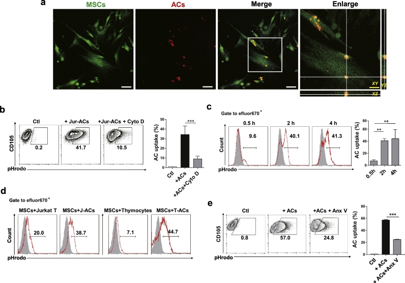
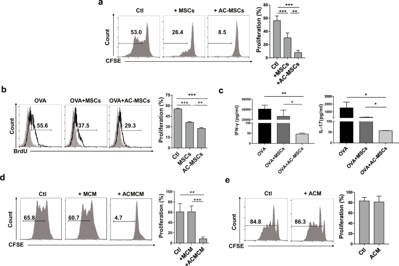
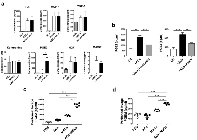
Human umbilical cord (UC) MSCs were co-cultured with ACs, and the engulfment of ACs by MSCs was either detected by flow cytometry or observed under confocal laser scanning microscope. Peripheral blood mononuclear cells (PBMCs) from healthy controls (HCs) were cultured in MSC conditioned medium (MCM) or MSC exposed to ACs (AC-MSC) conditioned medium (ACMCM), and then CD4 T cell proliferation was detected. Soluble factors including prostaglandin (PG)E2 in the supernatants of MSCs and AC-MSCs, as well as in the mouse peritoneal lavage fluids (PLF) were determined by enzyme-linked immunosorbent assay (ELISA). Cyclooxygenase (COX)2 inhibitors and siRNA transfection were utilized to determine the function of COX2/PGE2 in AC-MSC-mediated immunosuppression. PGE2 metabolites (PGEM) in the plasma of SLE patients were measured before and 24 h after MSC transplantation respectively.
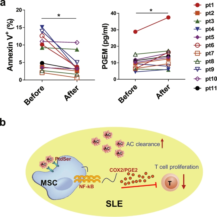
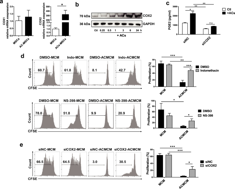
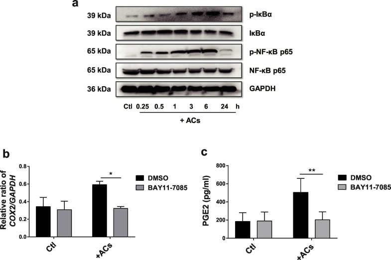
Human UC MSCs possessed the ability to engulf ACs. AC-MSCs increased MSC-mediated suppression of CD4 T cell proliferation compared to MSCs alone. Mechanistically, ACs stimulated MSCs to express COX2 and consequently produced PGE2 that inhibited T cell responses. NF-κB signalling pathway mediated the activation of COX2/PGE2 in AC-MSCs. Importantly, in patients with SLE, the plasma PGEM levels increased significantly in those with reduced apoptotic mononuclear cells in peripheral blood after MSC transplantation.
Clearance of ACs by MSCs contributes to immunosuppressive function via increasing PGE2 production. These findings reveal a previously unrecognized role of MSC-mediated phagocytosis of ACs in MSC-based immunotherapy. FUND: This study was supported by grants from the Chinese Major International (Regional) Joint Research Project (No. 81720108020), the Jiangsu Province Major Research and Development Program (No. BE2015602) and the Jiangsu Province 333 Talent Grant (BRA2016001). WJ. Chen was supported by the Intramural Research Program of NIH, NIDCR.
凋亡细胞(ACs)清除缺陷被认为与系统性红斑狼疮(SLE)的发病机制有关。间充质干细胞(MSCs)在治疗 SLE 方面显示出有前景的治疗效果,但 MSCs 是否吞噬 ACs 并有助于治疗 SLE 的潜在机制尚不清楚。
将人脐带(UC)MSCs 与 ACs 共培养,通过流式细胞术或共聚焦激光扫描显微镜观察 MSCs 吞噬 ACs 的情况。将健康对照(HC)的外周血单个核细胞(PBMCs)在 MSC 条件培养基(MCM)或暴露于 AC 的 MSC 条件培养基(ACMCM)中培养,然后检测 CD4 T 细胞增殖。通过酶联免疫吸附试验(ELISA)测定 MSCs 和 AC-MSCs 上清液以及小鼠腹腔灌洗液(PLF)中包括前列腺素(PG)E2 在内的可溶性因子。利用环氧化酶(COX)2 抑制剂和 siRNA 转染来确定 COX2/PGE2 在 AC-MSC 介导的免疫抑制中的作用。分别在 MSC 移植前和移植后 24 小时测量 SLE 患者血浆中的 PGE2 代谢物(PGEM)。
人 UC MSCs 具有吞噬 ACs 的能力。与单独的 MSC 相比,AC-MSCs 增加了 MSC 介导的 CD4 T 细胞增殖抑制作用。从机制上讲,AC 刺激 MSC 表达 COX2,并由此产生抑制 T 细胞反应的 PGE2。NF-κB 信号通路介导了 AC-MSCs 中 COX2/PGE2 的激活。重要的是,在 SLE 患者中,MSC 移植后外周血中凋亡单核细胞减少的患者,其血浆 PGEM 水平显著升高。
MSCs 通过增加 PGE2 的产生来清除 ACs,从而发挥免疫抑制功能。这些发现揭示了 MSC 吞噬 ACs 在 MSC 为基础的免疫治疗中的一个以前未被认识的作用。资助:本研究得到中国重大国际(地区)合作研究项目(No. 81720108020)、江苏省重大研发项目(No. BE2015602)和江苏省 333 人才项目(BRA2016001)的资助。WJ. Chen 得到 NIH、NIDCR 院内研究计划的支持。