Fei Honghua, Shi Mingyan, Chen Lianhong, Wang Zhe, Suo Lihua
Department of Endocrinology, People's Hospital of Rizhao, Rizhao, Shandong 276800, P.R. China.
Department of Blood Transfusion, Yantaishan Hospital, Yantai, Shandong 264000, P.R. China.
Exp Ther Med. 2019 Jul;18(1):389-396. doi: 10.3892/etm.2019.7527. Epub 2019 Apr 24.
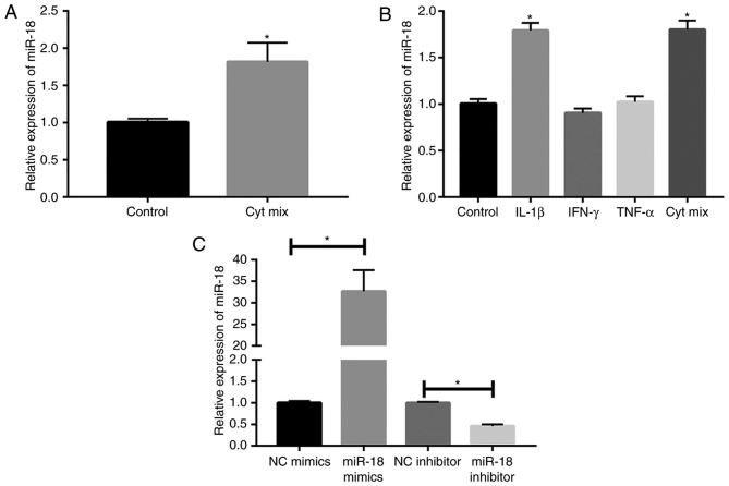
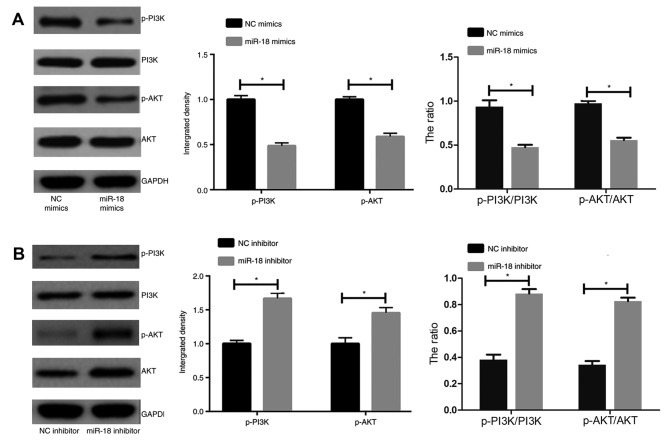
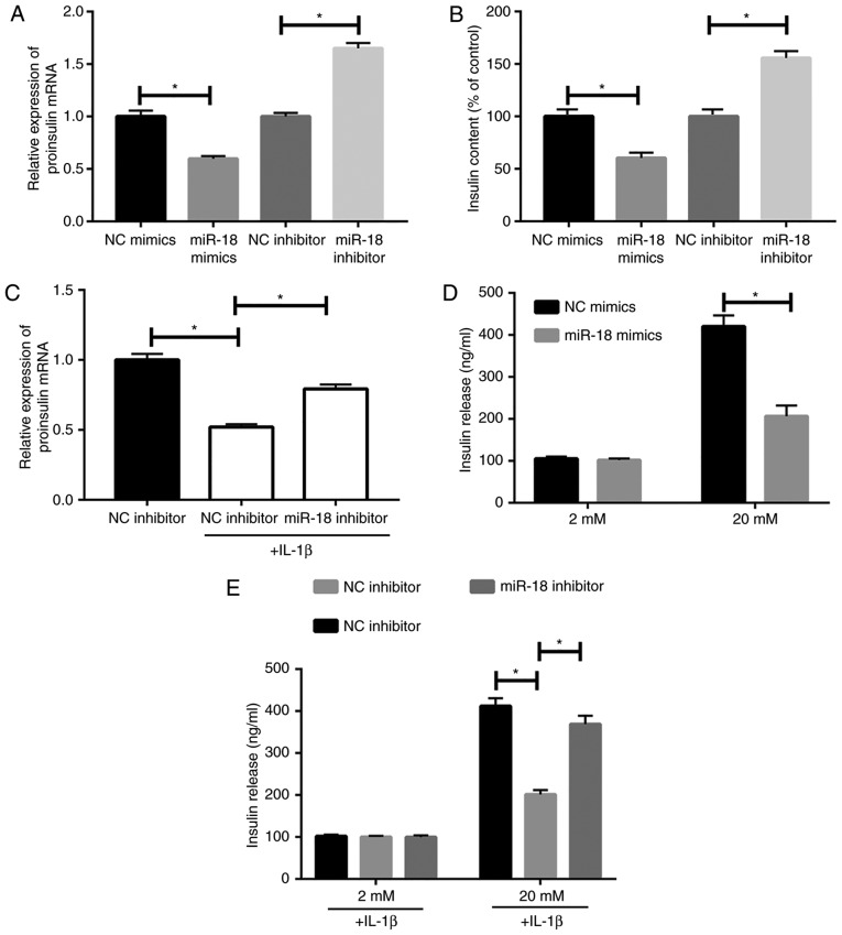
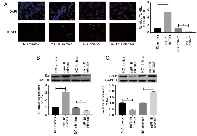
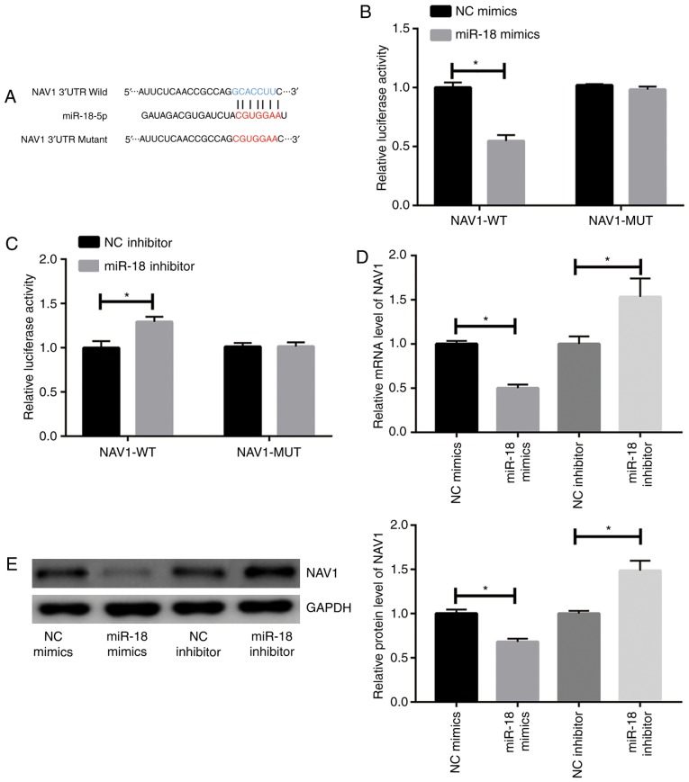
The detailed pathogenesis of diabetes mellitus (DM) remains to be fully elucidated. The purpose of the present study was to explore the role of microRNA (miR)-18 in DM and its underlying mechanisms, providing novel ideas for the treatment of the disease. After inflammatory factor-mediated induction, miR-18 expression in the islet β-cell line MIN6 was detected by reverse transcription-quantitative polymerase chain reaction (RT-qPCR). miR-18 mimics and miR-18 inhibitor were then constructed and transfected into MIN6 cells. The mRNA levels of pro-insulin in MIN6 cells were also detected by RT-qPCR. Released insulin levels and insulin secretion function of MIN6 cells were accessed by ELISA and glucose-stimulated insulin secretion assay, respectively. Apoptosis of MIN6 cells was detected by a terminal deoxynucleotidyl transferase-mediated deoxyuridinetriphosphate nick end labeling assay and western blot analysis of apoptotic proteins. The binding interaction of miR-18 and neuron navigator 1(NAV1), a constituent of the phosphoinositide 3-kinase (PI3K)/AKT pathway, was assessed using a dual-luciferase reporter gene assay. Finally, the regulatory effect of miR-18 on the PI3K/AKT pathway was determined by western blot analysis. After induction of inflammatory factors in MIN6 cells, miR-18 expression was markedly upregulated. Transfection with miR-18 mimics inhibited pro-insulin levels, as well as insulin production and secretion capacity. miR-18 knockdown partially abrogated the inhibited insulin secretion capacity induced by interleukin-1β (IL-1β) treatment. In addition, apoptosis of MIN6 cells was increased by miR-18 mimics. The dual-luciferase reporter gene assay confirmed the direct binding of miR-18 to NAV1. Western blot analysis suggested that miR-18 markedly inhibited the PI3K/AKT pathway in MIN6 cells. In conclusion, miR-18 expression is upregulated by IL-1β induction in islet β-cells. It was demonstrated that miR-18 promotes apoptosis of islet β-cells at least partially by inhibiting NAV1 expression and insulin production via suppression of the PI3K/AKT pathway.
糖尿病(DM)的详细发病机制仍有待充分阐明。本研究的目的是探讨微小RNA(miR)-18在糖尿病中的作用及其潜在机制,为该疾病的治疗提供新思路。在炎症因子介导的诱导后,通过逆转录-定量聚合酶链反应(RT-qPCR)检测胰岛β细胞系MIN6中miR-18的表达。然后构建miR-18模拟物和miR-18抑制剂并转染到MIN6细胞中。也通过RT-qPCR检测MIN6细胞中胰岛素原的mRNA水平。分别通过ELISA和葡萄糖刺激的胰岛素分泌试验评估MIN6细胞释放的胰岛素水平和胰岛素分泌功能。通过末端脱氧核苷酸转移酶介导的脱氧尿苷三磷酸缺口末端标记试验和凋亡蛋白的蛋白质印迹分析检测MIN6细胞的凋亡。使用双荧光素酶报告基因试验评估miR-18与磷酸肌醇3-激酶(PI3K)/AKT途径的组成部分神经元导航蛋白1(NAV1)的结合相互作用。最后,通过蛋白质印迹分析确定miR-18对PI3K/AKT途径的调节作用。在MIN6细胞中诱导炎症因子后,miR-18表达明显上调。用miR-18模拟物转染抑制了胰岛素原水平以及胰岛素的产生和分泌能力。miR-18敲低部分消除了白细胞介素-1β(IL-1β)处理诱导的胰岛素分泌能力抑制。此外,miR-18模拟物增加了MIN6细胞的凋亡。双荧光素酶报告基因试验证实了miR-18与NAV1的直接结合。蛋白质印迹分析表明,miR-18显著抑制MIN6细胞中的PI3K/AKT途径。总之,胰岛β细胞中IL-1β诱导上调miR-18表达。结果表明,miR-18至少部分通过抑制NAV1表达和经由抑制PI3K/AKT途径的胰岛素产生来促进胰岛β细胞凋亡。