Bandeira Janete Shatkoski, Antunes Luciana da Conceição, Soldatelli Matheus Dorigatti, Sato João Ricardo, Fregni Felipe, Caumo Wolnei
Laboratory of Pain and Neuromodulation, Universidade Federal do Rio Grande do Sul (UFRGS), Porto Alegre, Brazil.
Department of Nutrition, Health Science Center, Universidade Federal de Santa Catarina (UFSC), Florianópolis, Brazil.
Front Hum Neurosci. 2019 Jun 13;13:200. doi: 10.3389/fnhum.2019.00200. eCollection 2019.
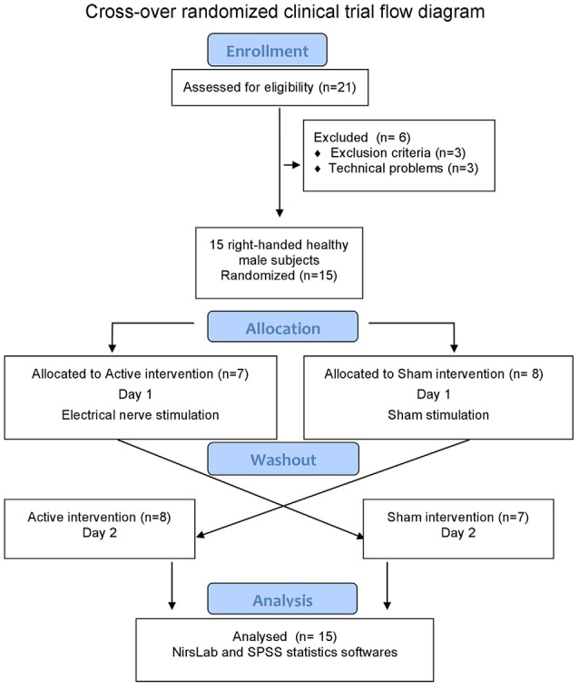
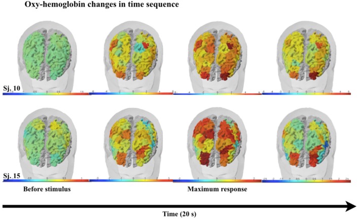
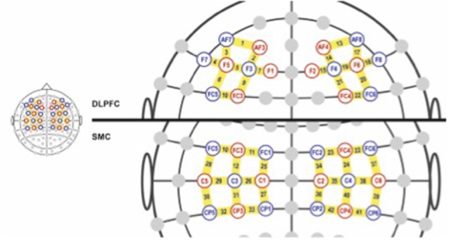
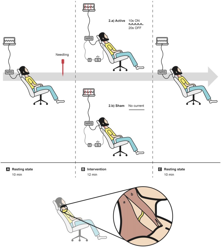
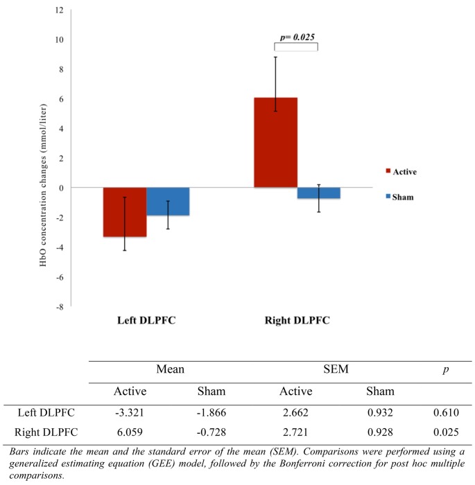
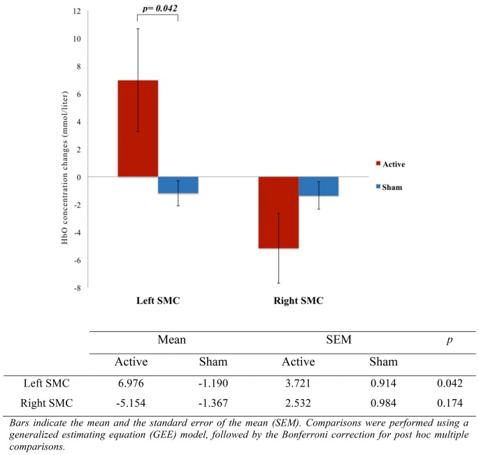
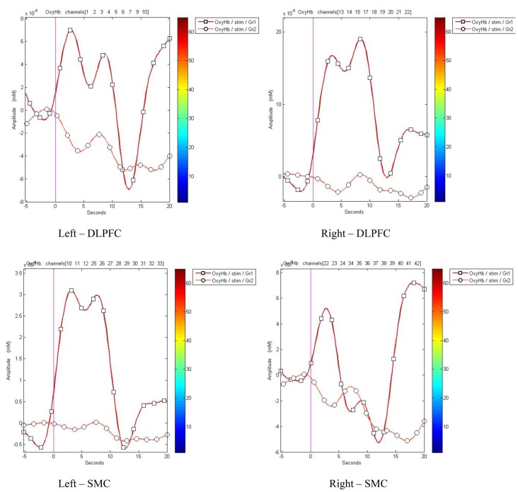
Peripheral electrical stimulation (PES), which encompasses several techniques with heterogeneous physiological responses, has shown in some cases remarkable outcomes for pain treatment and clinical rehabilitation. However, results are still mixed, mainly because there is a lack of understanding regarding its neural mechanisms of action. In this study, we aimed to assess its effects by measuring cortical activation as indexed by functional near infrared spectroscopy (fNIRS). fNIRS is a functional optical imaging method to evaluate hemodynamic changes in oxygenated (HbO) and de-oxygenated (HbR) blood hemoglobin concentrations in cortical capillary networks that can be related to cortical activity. We hypothesized that non-painful PES of accessory spinal nerve (ASN) can promote cortical activation of sensorimotor cortex (SMC) and dorsolateral prefrontal cortex (DLPFC) pain processing cortical areas. Fifteen healthy volunteers received both active and sham ASN electrical stimulation in a crossover study. The hemodynamic cortical response to unilateral right ASN burst electrical stimulation with 10 Hz was measured by a 40-channel fNIRS system. The effect of ASN electrical stimulation over HbO concentration in cortical areas of interest (CAI) was observed through the activation of right-DLPFC ( = 0.025) and left-SMC ( = 0.042) in the active group but not in sham group. Regarding left-DLPFC ( = 0.610) and right-SMC ( = 0.174) there was no statistical difference between groups. As in non-invasive brain stimulation (NIBS) top-down modulation, bottom-up electrical stimulation to the ASN seems to activate the same critical cortical areas on pain pathways related to sensory-discriminative and affective-motivational pain dimensions. These results provide additional mechanistic evidence to develop and optimize the use of peripheral nerve electrical stimulation as a neuromodulatory tool (NCT03295370- www.clinicaltrials.gov).
外周电刺激(PES)涵盖了多种具有不同生理反应的技术,在某些情况下已显示出在疼痛治疗和临床康复方面的显著效果。然而,结果仍然参差不齐,主要是因为对其神经作用机制缺乏了解。在本研究中,我们旨在通过测量功能性近红外光谱(fNIRS)所索引的皮质激活来评估其效果。fNIRS是一种功能性光学成像方法,用于评估皮质毛细血管网络中氧合血红蛋白(HbO)和脱氧血红蛋白(HbR)浓度的血流动力学变化,这些变化可能与皮质活动有关。我们假设副脊神经(ASN)的非疼痛性PES可以促进感觉运动皮层(SMC)和背外侧前额叶皮层(DLPFC)疼痛处理皮质区域的皮质激活。在一项交叉研究中,15名健康志愿者接受了主动和假ASN电刺激。通过40通道fNIRS系统测量对10 Hz单侧右ASN爆发性电刺激的血流动力学皮质反应。在主动组中观察到ASN电刺激对感兴趣皮质区域(CAI)中HbO浓度的影响,表现为右侧DLPFC(P = 0.025)和左侧SMC(P = 0.042)的激活,而假刺激组未观察到。关于左侧DLPFC(P = 0.610)和右侧SMC(P = 0.174),两组之间没有统计学差异。与非侵入性脑刺激(NIBS)的自上而下调节一样,对ASN的自下而上电刺激似乎激活了与感觉辨别和情感动机性疼痛维度相关的疼痛通路中的相同关键皮质区域。这些结果为开发和优化将外周神经电刺激作为一种神经调节工具提供了额外的机制证据(NCT03295370 - www.clinicaltrials.gov)。