Department of Radiology and Nuclear Medicine (766), Radboud university medical center, P.O. Box 9101, Nijmegen, The Netherlands.
Erwin L Hahn Institute for Magnetic Resonance Imaging, UNESCO World Cultural, Heritage Zollverein, Kokereiallee 7, Building C84, D-45141, Essen, Germany.
Med Phys. 2019 Sep;46(9):3893-3905. doi: 10.1002/mp.13696. Epub 2019 Aug 1.
In vivo H and P magnetic resonance spectroscopic imaging (MRSI) provide complementary information on the biology of prostate cancer. In this work we demonstrate the feasibility of performing multiparametric imaging (mpMRI) and H and P spectroscopic imaging of the prostate using a P and H endorectal radiofrequency coil (ERC) in combination with a multitransmit body array at 7 Tesla (T).
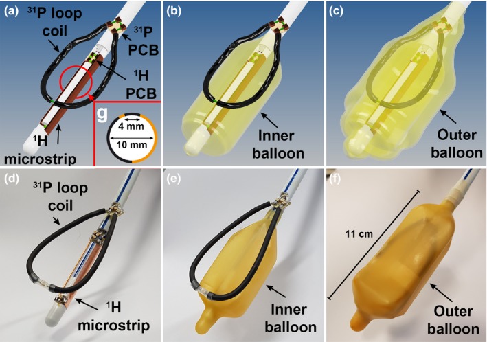
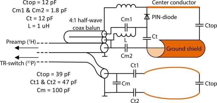
An ERC with a P transceiver loop coil and H receive (Rx) asymmetric microstrip ( P/ H ERC) was designed, constructed and tested in combination with an external 8-channel H transceiver body array coil (8CH). Electromagnetic field simulations and measurements and in vivo temperature measurements of the ERC were performed for safety validation. In addition, the signal-to-noise (SNR) benefit of the H microstrip with respect to the 8CH was evaluated. Finally, the feasibility of the setup was tested in one volunteer and three patients with prostate cancer by performing T -weighted and diffusion-weighted imaging in combination with H and P spectroscopic imaging.
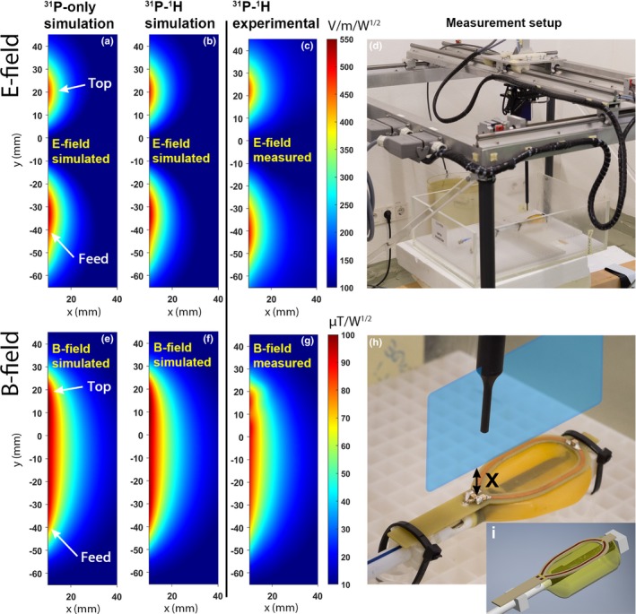
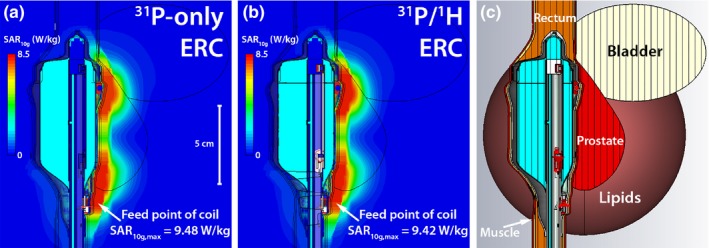
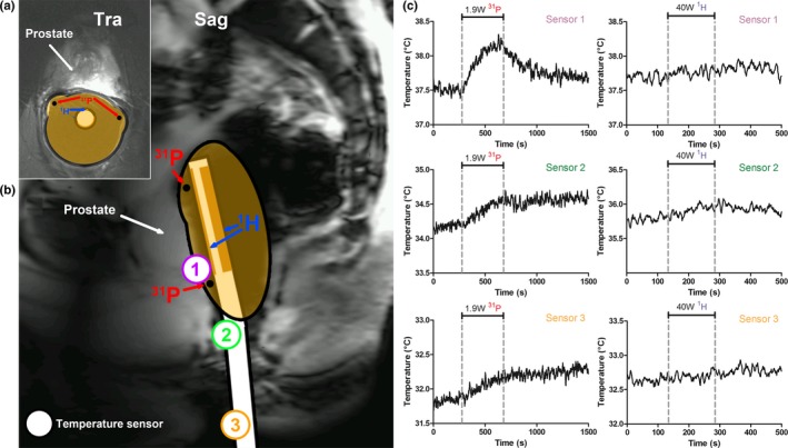
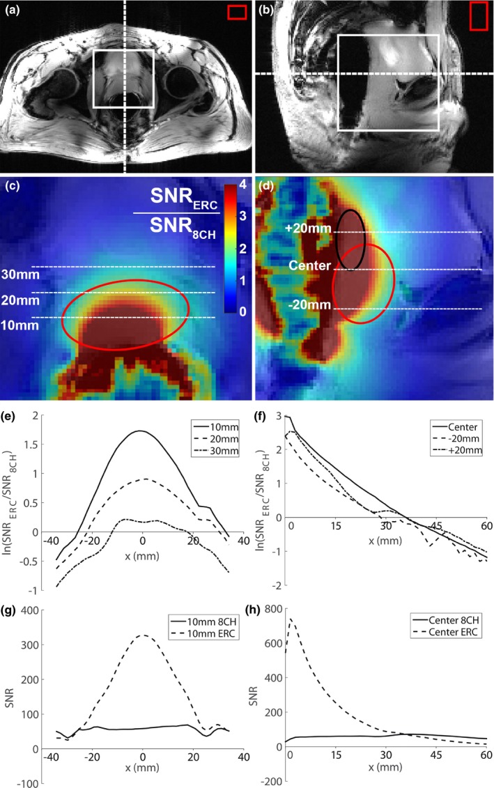
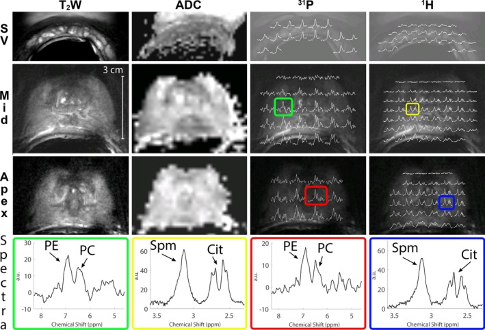
Electromagnetic field simulations of the P loop coil showed no differences in the E- and B-fields of the P/ H ERC compared with a previously safety validated ERC without H microstrip. The hotspot of the specific absorption rate (SAR) at the feed point of the P/ H ERC loop coil was 9.42 W/kg when transmitting on P at 1 W. Additional in vivo measurements showed a maximum temperature increase at the SAR hotspot of 0.7°C over 6 min on P at 1.9 W transmit (Tx) power, indicating safe maximum power levels. When transmitting with the external H body array at 40W for 2:30 min, the temperature increase around the ERC was < 0.3°C. Up to 3.5 cm into the prostate the H microstrip of the ERC provided higher SNR than the 8CH. The total coil combination allowed acquisition of an mpMRI protocol and the assessment of P and H metabolites of the prostate in all test subjects.
We developed a setup with a P transceiver and H Rx endorectal coil in combination with an 8-channel transceiver external body array coil and demonstrated its safety and feasibility for obtaining multiparametric imaging and H and P MRSI at 7T in patients with prostate cancer within one MR examination.
体内 H 和 P 磁共振波谱成像(MRSI)提供了前列腺癌生物学的互补信息。在这项工作中,我们展示了在 7 特斯拉(T)下使用直肠内 P 和 H 射频线圈(ERC)与多发射体阵列结合进行前列腺多参数成像(mpMRI)和 H 和 P 波谱成像的可行性。
设计、构建并测试了一种带有 P 收发器环形线圈和 H 接收(Rx)非对称微带(P/H ERC)的 ERC,并与外部 8 通道 H 收发体阵列线圈(8CH)结合使用。进行了 ERC 的电磁场模拟和测量以及体内温度测量,以验证安全性。此外,还评估了 H 微带相对于 8CH 的信噪比(SNR)优势。最后,在一名志愿者和三名前列腺癌患者中测试了该设备的可行性,通过 T 加权和扩散加权成像以及 H 和 P 波谱成像进行。
P 环形线圈的电磁场模拟显示,与没有 H 微带的先前经过安全验证的 ERC 相比,P/H ERC 的 E 场和 B 场没有差异。当在 1 W 时在 P 上发送时,P/H ERC 环形线圈的比吸收率(SAR)热点处的 SAR 为 9.42 W/kg。额外的体内测量显示,在 1.9 W 发射(Tx)功率下,P 上 6 分钟时 SAR 热点处的最大温度升高为 0.7°C,表明安全的最大功率水平。当在 40W 下以 2:30 分钟的时间通过外部 H 体阵列传输时,ERC 周围的温度升高<0.3°C。在进入前列腺 3.5 厘米的范围内,ERC 的 H 微带比 8CH 提供了更高的 SNR。总的线圈组合允许采集多参数成像协议,并在所有测试对象中评估前列腺的 P 和 H 代谢物。
我们开发了一种带有 P 收发器和 H 直肠内 Rx 线圈的设备,与外部 8 通道收发体阵列线圈相结合,并在一名前列腺癌患者中展示了其在单次磁共振检查中获得多参数成像和 H 和 P MRSI 的安全性和可行性。