Division of Radiology and Nuclear Medicine, Department of Surgical Sciences, Uppsala University, Uppsala, Sweden.
Medical Physics, Uppsala University Hospital, Uppsala, Sweden.
Theranostics. 2019 May 25;9(12):3476-3484. doi: 10.7150/thno.31970. eCollection 2019.
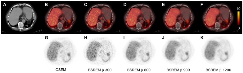
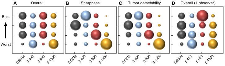
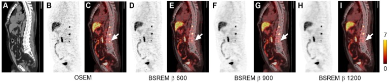
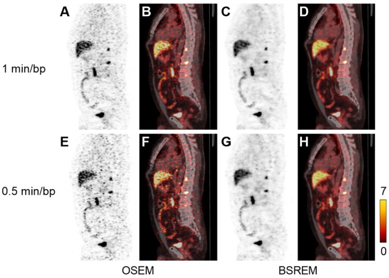
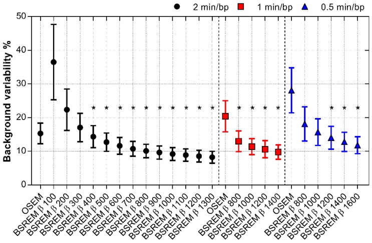
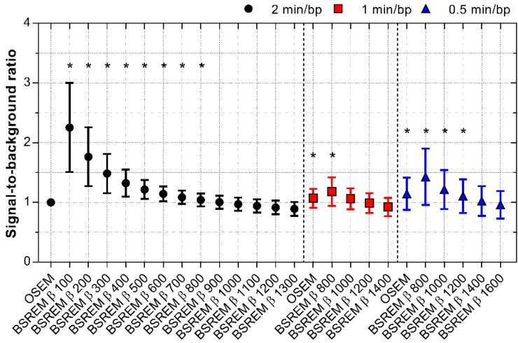
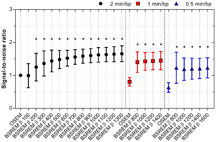
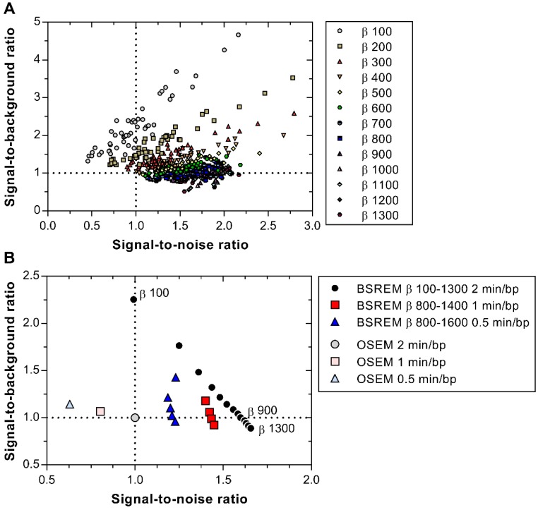
Accurate localization of recurrent prostate cancer (PCa) is critical, especially if curative therapy is intended. With the aim to optimize target-to-background uptake ratio in Ga-PSMA-11 PET, we investigated the image quality and quantitative measures of regularized reconstruction by block-sequential regularized expectation maximization (BSREM). The study encompassed retrospective reconstruction and analysis of 20 digital time-of-flight (TOF) PET/CT examinations acquired 60 min post injection of 2 MBq/kg of Ga-PSMA-11 in PCa patients with biochemical relapse after primary treatment. Reconstruction by ordered-subsets expectation maximization (OSEM; 3 iterations, 16 subsets, 5 mm gaussian postprocessing filter) and BSREM (β-values of 100-1600) were used, both including TOF and point spread function (PSF) recovery. Background variability (BV) was measured by placing a spherical volume of interest in the right liver lobe and defined as the standard deviation divided by the mean standardized uptake value (SUV). The image quality was evaluated in terms of signal-to-noise ratio (SNR) and signal-to-background ratio (SBR), using SUV of the lesions. A visual assessment was performed by four observers. OSEM reconstruction produced images with a BV of 15%, whereas BSREM with a β-value above 300 resulted in lower BVs than OSEM (36% with β 100, 8% with β 1300). Decreasing the acquisition duration from 2 to 1 and 0.5 min per bed position increased BV for both reconstruction methods, although BSREM with β-values equal to or higher than 800 and 1200, respectively, kept the BV below 15%. In comparison of BSREM with OSEM, the mean SNR improved by 25 to 66% with an increasing β-value in the range of 200-1300, whereas the mean SBR decreased with an increasing β-value, ranging from 0 to 125% with a β-value of 100 and 900, respectively. Decreased acquisition duration resulted in β-values of 800 to 1000 and 1200 to 1400 for 1 and 0.5 min per bed position, respectively, producing improved image quality measures compared with OSEM at a full acquisition duration of 2 min per bed position. The observer study showed a slight overall preference for BSREM β 900 although the interobserver variability was high. BSREM image reconstruction with β-values in the range of 400-900 resulted in lower BV and similar or improved SNR and SBR in comparison with OSEM.
准确定位复发性前列腺癌(PCa)至关重要,特别是如果计划进行治愈性治疗。为了优化 Ga-PSMA-11 PET 中的靶标与背景摄取比,我们研究了正则化期望最大化的块顺序正则化(BSREM)的图像质量和定量测量。该研究包括回顾性重建和分析 20 例数字时间飞行(TOF)PET/CT 检查,这些检查在原发性治疗后生化复发的 PCa 患者中在 60 分钟时注射了 2 MBq/kg 的 Ga-PSMA-11。使用有序子集期望最大化(OSEM;3 次迭代,16 个子集,5 毫米高斯后处理滤波器)和 BSREM(β 值为 100-1600)进行重建,两者均包括 TOF 和点扩散函数(PSF)恢复。通过在右肝叶放置一个球形感兴趣区来测量背景变异性(BV),并将其定义为标准偏差除以标准化摄取值(SUV)的平均值。使用病变的 SUV 评估图像质量,包括信噪比(SNR)和信号与背景比(SBR)。由四名观察者进行视觉评估。OSEM 重建产生的图像 BV 为 15%,而 BSREM 的β值大于 300 则产生低于 OSEM 的 BV(β值为 100 时为 36%,β值为 1300 时为 8%)。将采集时间从 2 分钟缩短到 1 分钟和 0.5 分钟/床位位置,两种重建方法的 BV 都会增加,但是 BSREM 的β值等于或高于 800 和 1200 时,BV 保持在 15%以下。与 OSEM 相比,BSREM 的平均 SNR 提高了 25%至 66%,β 值在 200-1300 范围内增加,而平均 SBR 随着β值的增加而降低,β 值为 100 时为 0 至 125%,β 值为 900 时为 900。对于 1 分钟和 0.5 分钟/床位位置,采集时间减少导致 BSREM 的β 值分别为 800 至 1000 和 1200 至 1400,与在 2 分钟/床位位置的完整采集时间相比,BSREM 产生了更好的图像质量测量值。观察者研究显示,尽管观察者间的变异性较高,但总体上对 BSREMβ900 有轻微偏好。与 OSEM 相比,BSREM 图像重建的β 值在 400-900 范围内可降低 BV,并在 SNR 和 SBR 方面具有相似或改善。