Laboratory of Lymphocyte Signaling & Molecular Imaging, MOE Key Laboratory of Protein Sciences, School of Life Sciences, Collaborative Innovation Center for Diagnosis and Treatment of Infectious Diseases, Institute for Immunology, Center for Life Sciences, Beijing Key Lab for Immunological Research on Chronic Diseases, Tsinghua University, Beijing, China.
School of Life Sciences, Tsinghua-Peking Joint Center for Life Sciences, Beijing Advanced Innovation Center for Structural Biology, Beijing Frontier Research Center for Biological Structure, Tsinghua University, Beijing, China.
Elife. 2019 Jul 10;8:e42271. doi: 10.7554/eLife.42271.
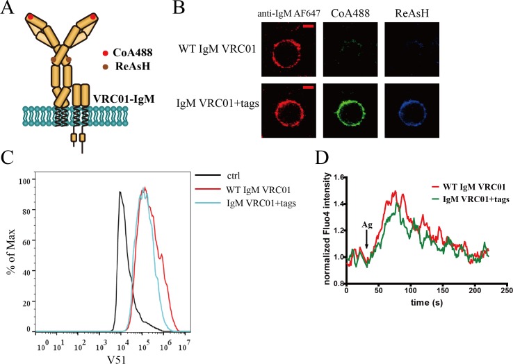
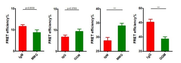
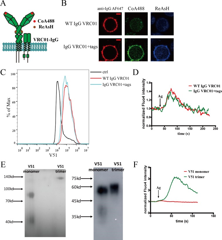
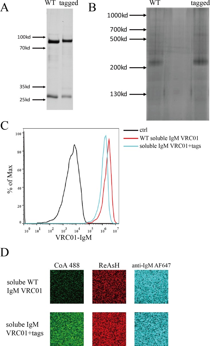
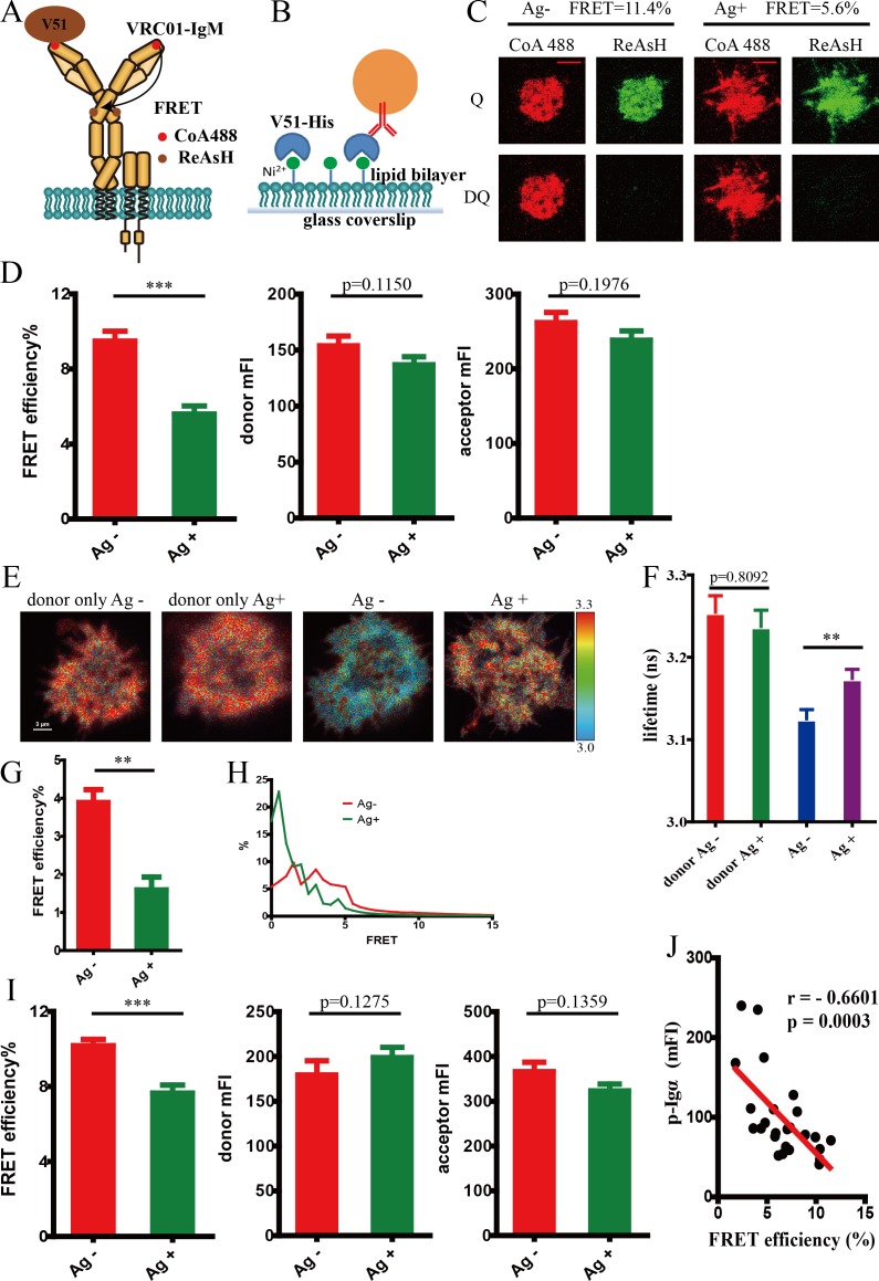
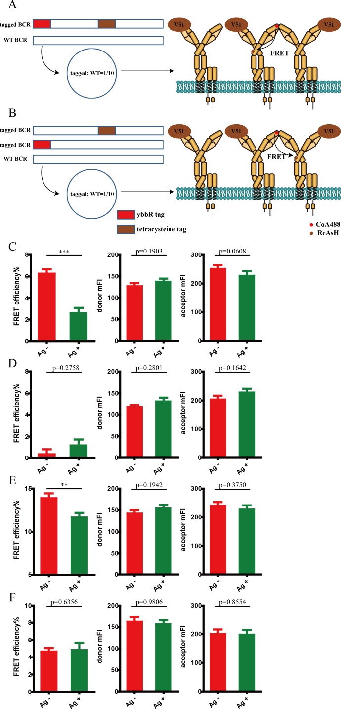
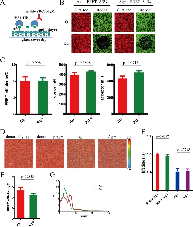
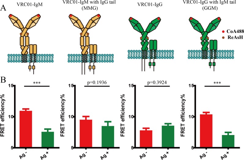
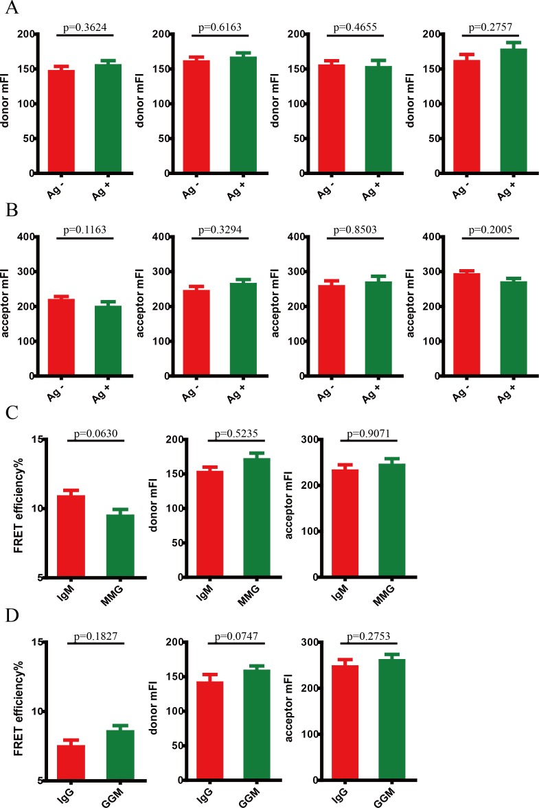
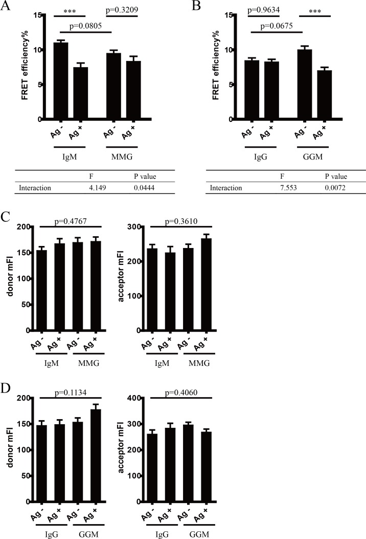
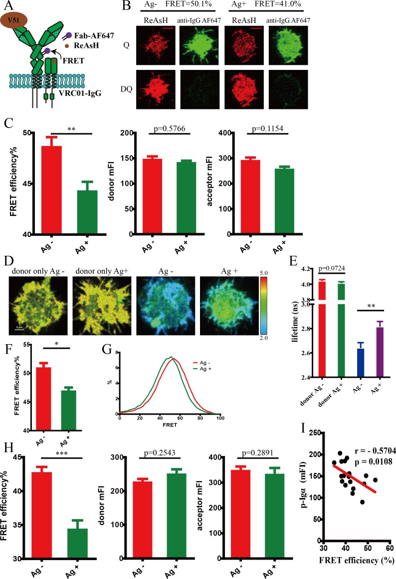
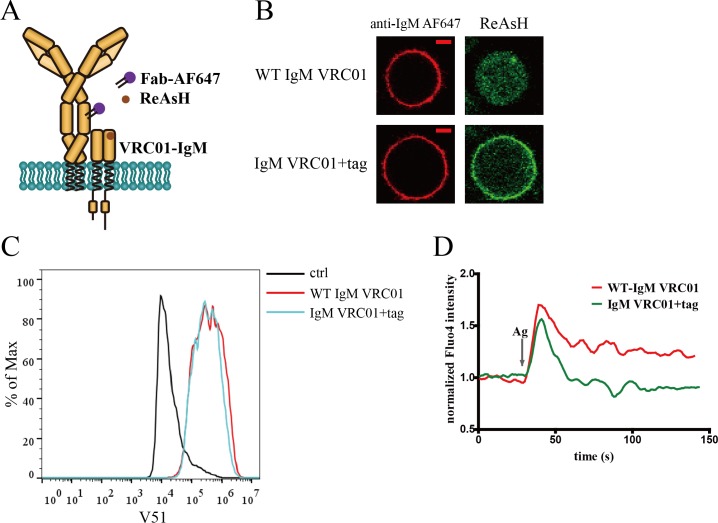
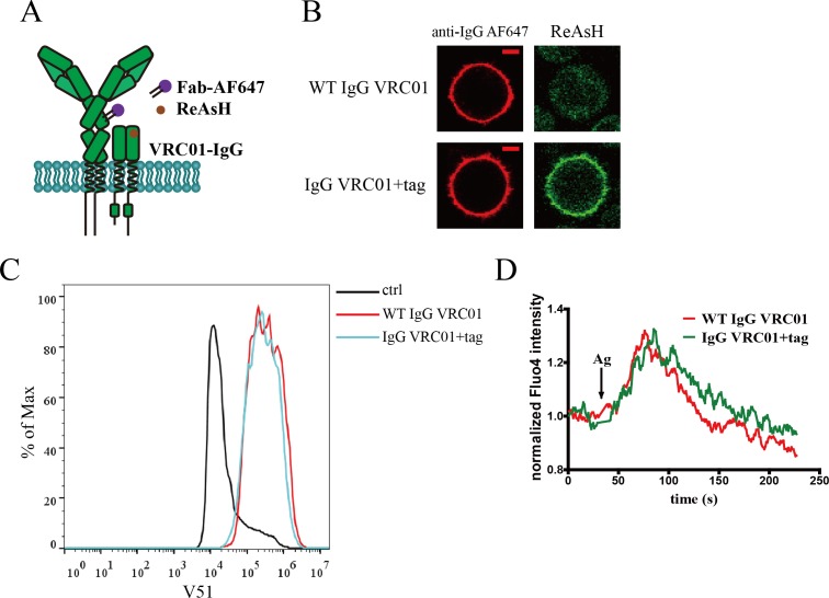
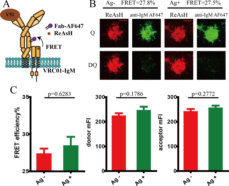
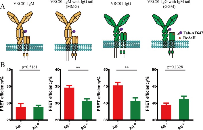
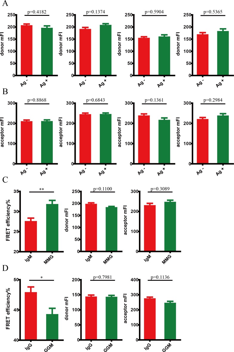
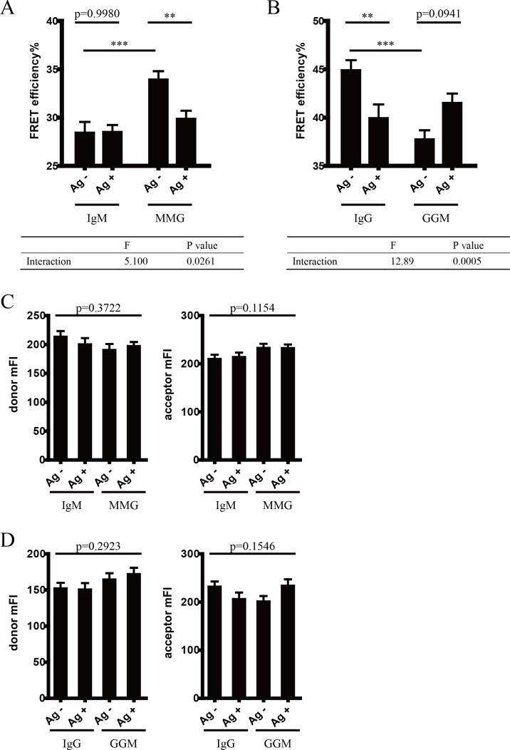
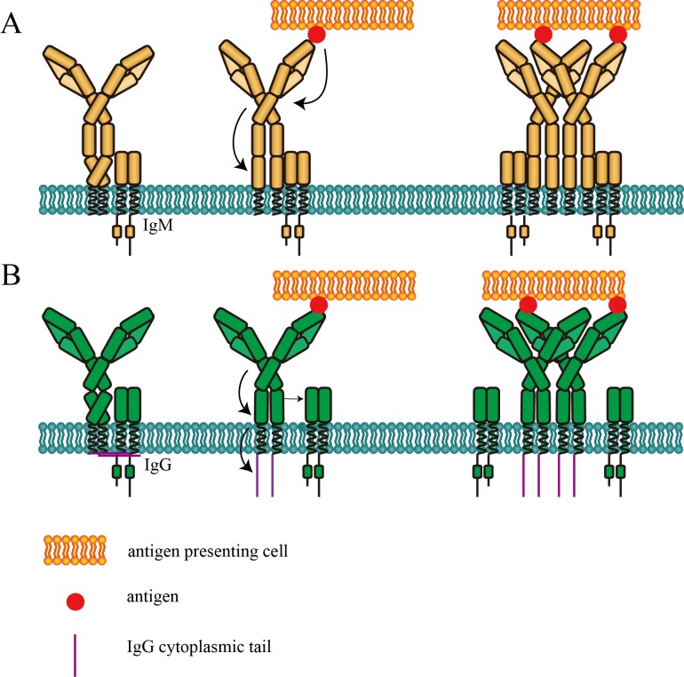
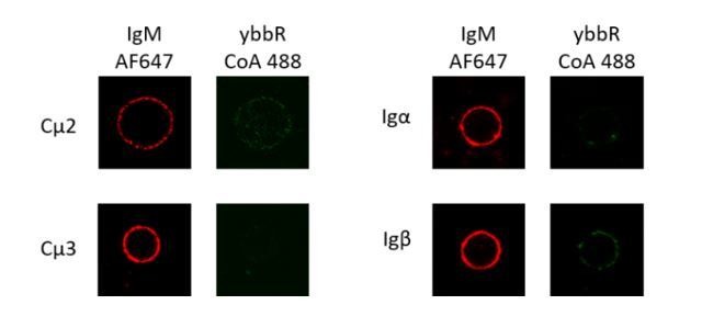
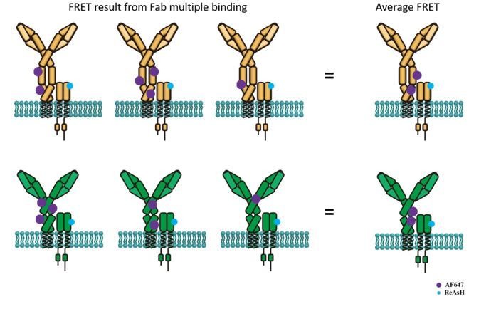
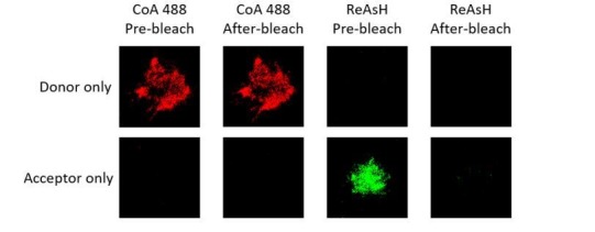
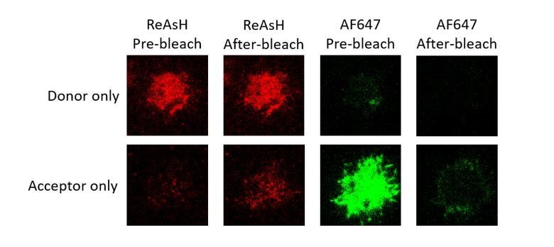
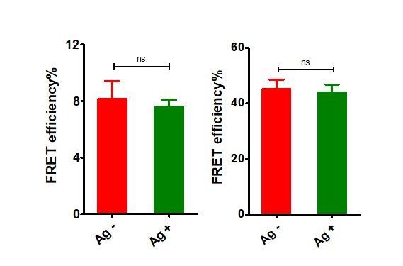
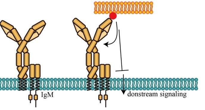
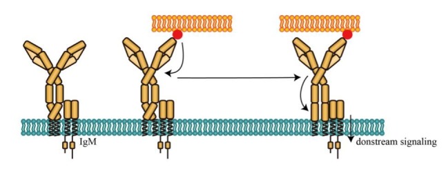
B lymphocytes use B cell receptors (BCRs) to recognize antigens. It is still not clear how BCR transduces antigen-specific physical signals upon binding across cell membrane for the conversion to chemical signals, triggering downstream signaling cascades. It is hypothesized that through a series of conformational changes within BCR, antigen engagement in the extracellular domain of BCR is transduced to its intracellular domain. By combining site-specific labeling methodology and FRET-based assay, we monitored conformational changes in the extracellular domains within BCR upon antigen engagement. Conformational changes within heavy chain of membrane-bound immunoglobulin (mIg), as well as conformational changes in the spatial relationship between mIg and Igβ were observed. These conformational changes were correlated with the strength of BCR activation and were distinct in IgM- and IgG-BCR. These findings provide molecular mechanisms to explain the fundamental aspects of BCR activation and a framework to investigate ligand-induced molecular events in immune receptors.
B 细胞利用 B 细胞受体 (BCR) 识别抗原。目前尚不清楚 BCR 在跨细胞膜结合后如何将抗原特异性物理信号转导为化学信号,从而触发下游信号级联。据推测,通过 BCR 内的一系列构象变化,BCR 细胞外结构域内的抗原结合被转导到其细胞内结构域。通过结合位点特异性标记方法和基于 FRET 的测定法,我们监测了抗原结合后 BCR 细胞外结构域内的构象变化。观察到膜结合免疫球蛋白 (mIg) 的重链内的构象变化,以及 mIg 和 Igβ 之间空间关系的构象变化。这些构象变化与 BCR 激活的强度相关,并且在 IgM 和 IgG-BCR 中是不同的。这些发现为解释 BCR 激活的基本方面提供了分子机制,并为研究免疫受体中配体诱导的分子事件提供了框架。