• 文献检索
  • 文档翻译
  • 深度研究
  • 学术资讯
  • Suppr Zotero 插件Zotero 插件
  • 邀请有礼
  • 套餐&价格
  • 历史记录
应用&插件
Suppr Zotero 插件Zotero 插件浏览器插件Mac 客户端Windows 客户端微信小程序
定价
高级版会员购买积分包购买API积分包
服务
文献检索文档翻译深度研究API 文档MCP 服务
关于我们
关于 Suppr公司介绍联系我们用户协议隐私条款
关注我们

Suppr 超能文献

核心技术专利:CN118964589B侵权必究
粤ICP备2023148730 号-1Suppr @ 2026

文献检索

告别复杂PubMed语法,用中文像聊天一样搜索,搜遍4000万医学文献。AI智能推荐,让科研检索更轻松。

立即免费搜索

文件翻译

保留排版,准确专业,支持PDF/Word/PPT等文件格式,支持 12+语言互译。

免费翻译文档

深度研究

AI帮你快速写综述,25分钟生成高质量综述,智能提取关键信息,辅助科研写作。

立即免费体验

线粒体膜重塑 GTP 酶 Mgm1 的结构与组装

Structure and assembly of the mitochondrial membrane remodelling GTPase Mgm1.

机构信息

Crystallography, Max-Delbrück-Centrum for Molecular Medicine, Berlin, Germany.

Department of Structural Biology, Max Planck Institute of Biophysics, Frankfurt am Main, Germany.

出版信息

Nature. 2019 Jul;571(7765):429-433. doi: 10.1038/s41586-019-1372-3. Epub 2019 Jul 10.

DOI:10.1038/s41586-019-1372-3
PMID:31292547
原文链接:https://pmc.ncbi.nlm.nih.gov/articles/PMC7116848/
Abstract

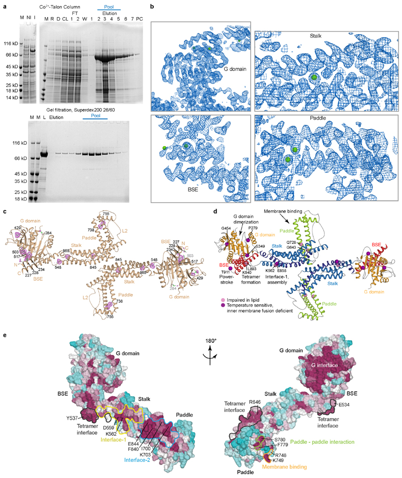
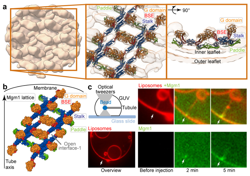
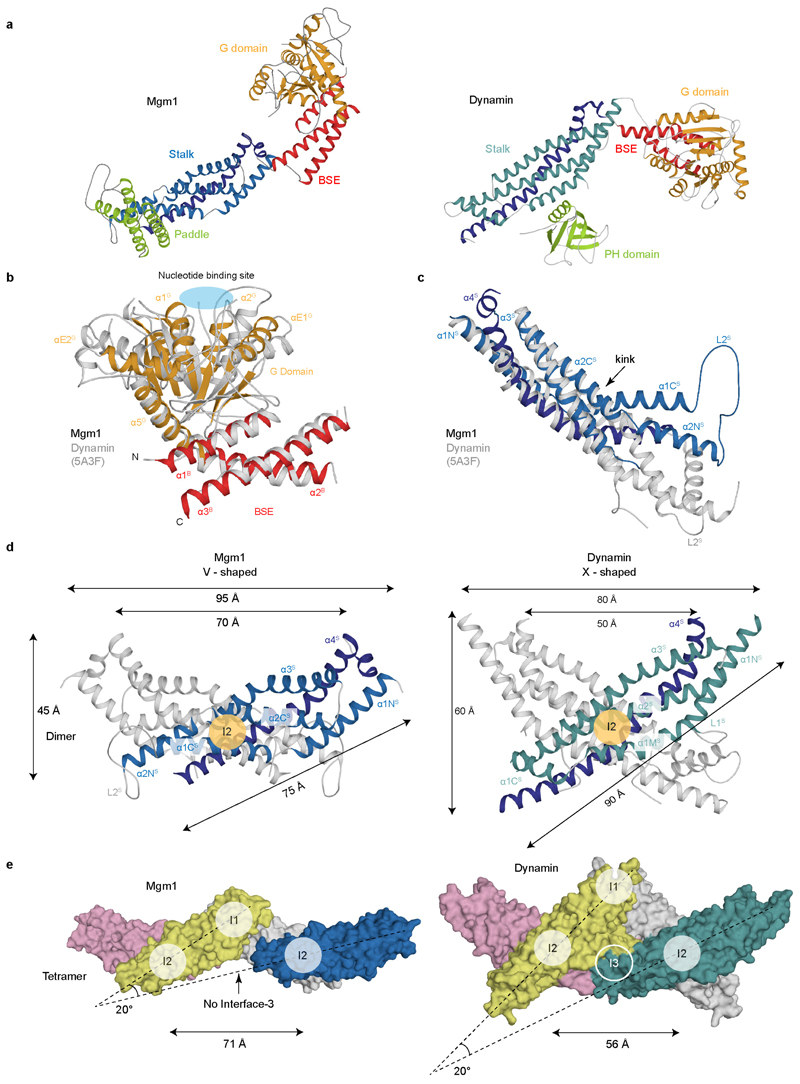
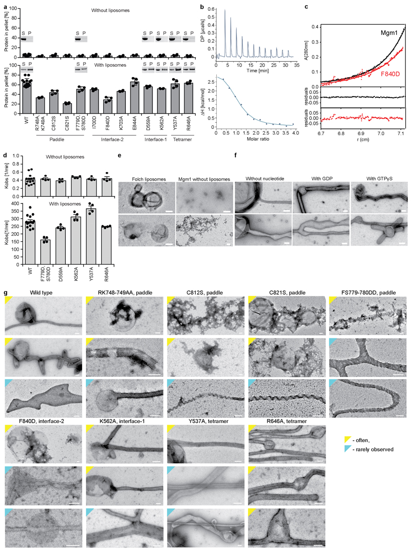
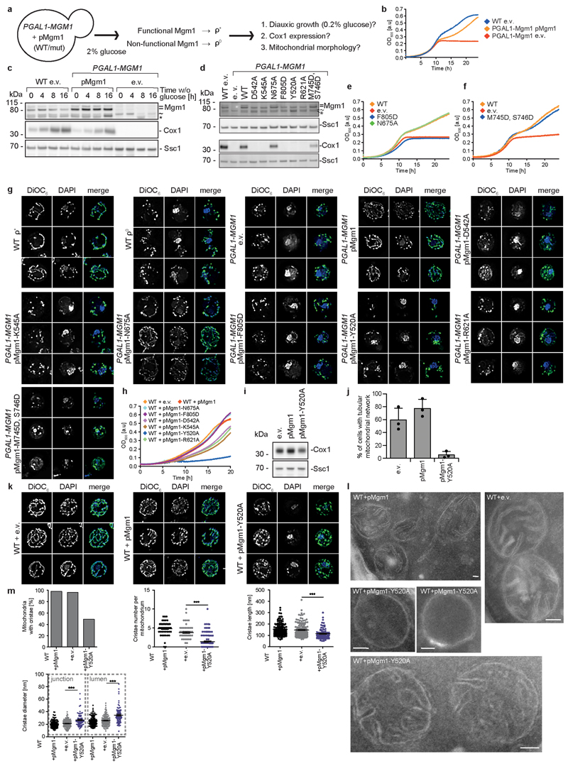
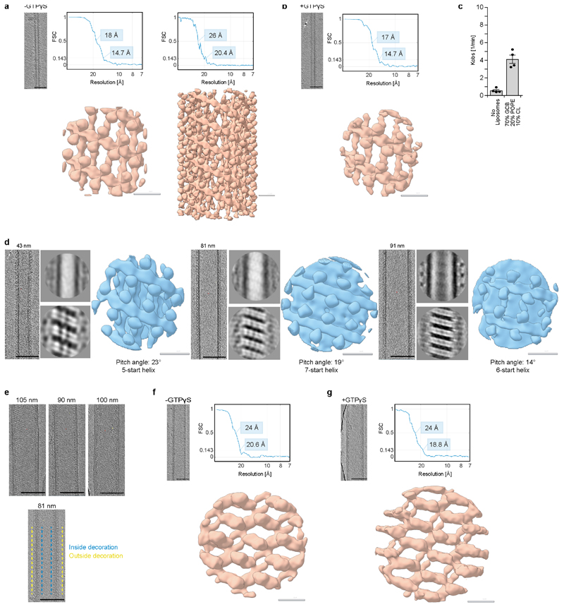
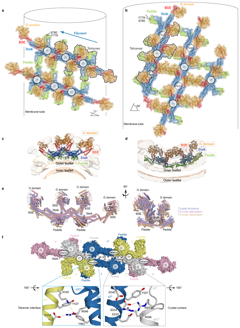
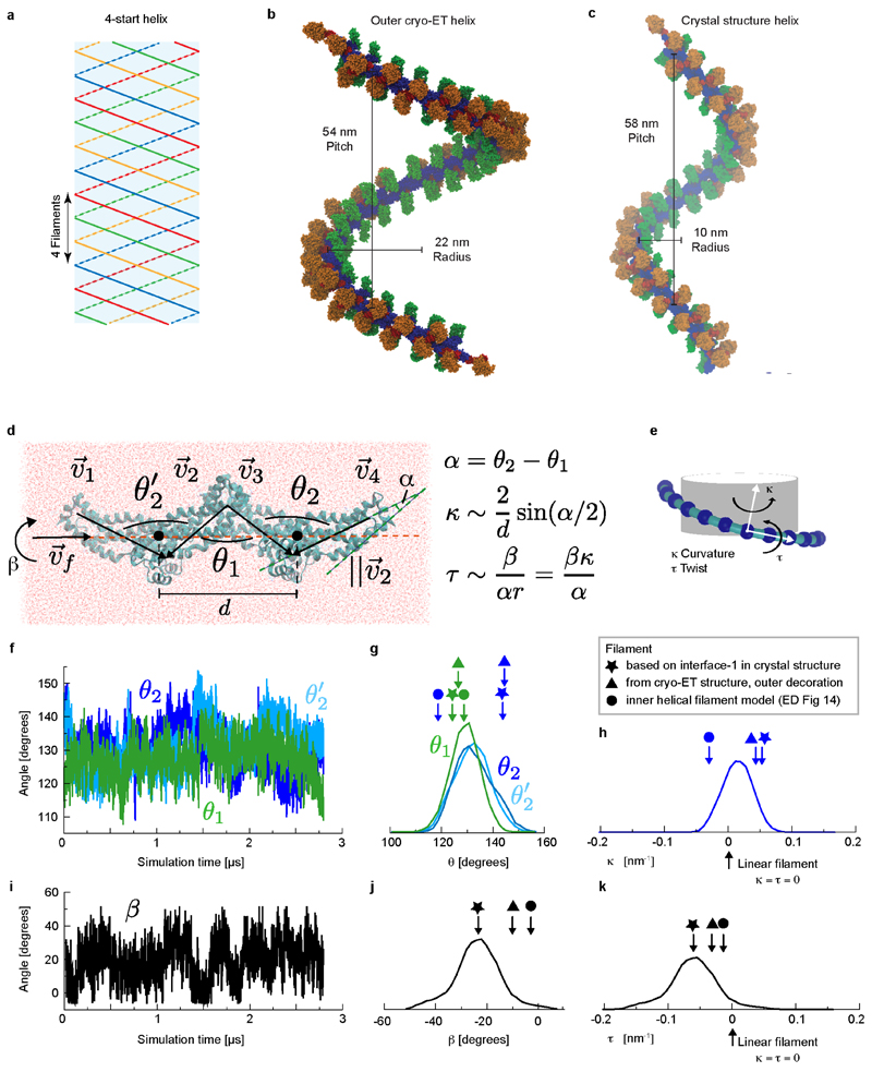
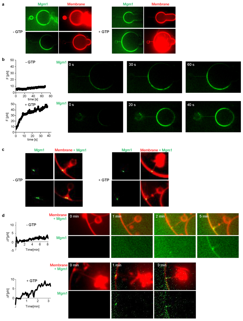
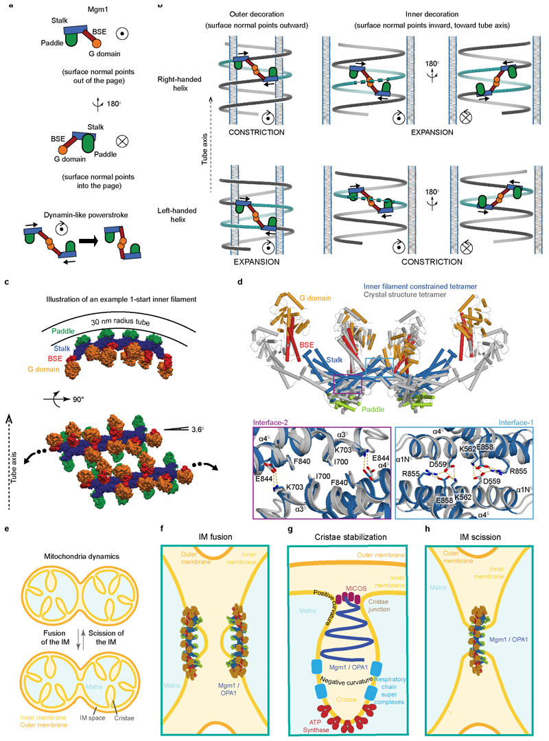
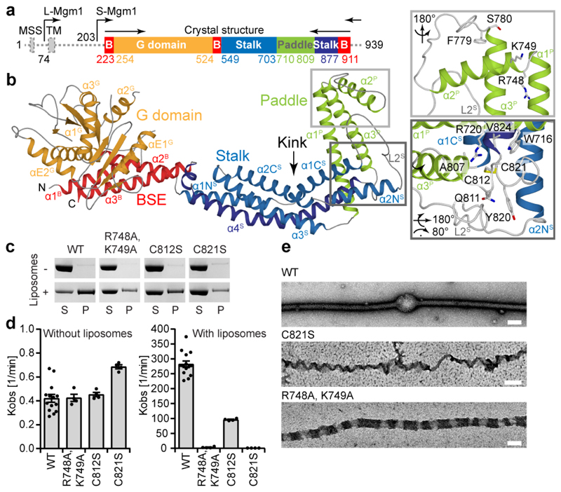
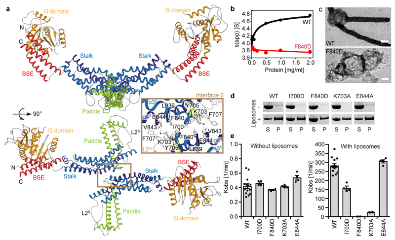
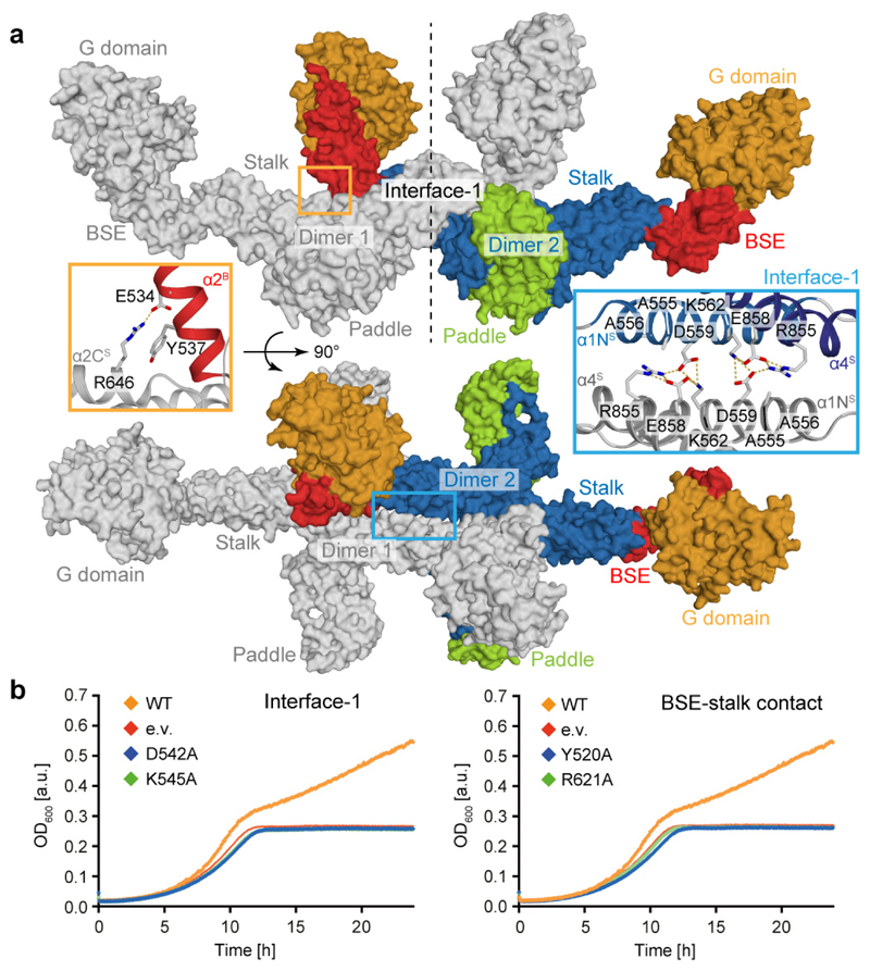
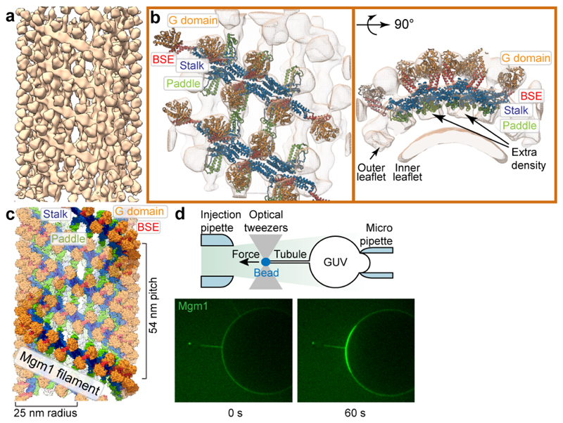
Balanced fusion and fission are key for the proper function and physiology of mitochondria. Remodelling of the mitochondrial inner membrane is mediated by the dynamin-like protein mitochondrial genome maintenance 1 (Mgm1) in fungi or the related protein optic atrophy 1 (OPA1) in animals. Mgm1 is required for the preservation of mitochondrial DNA in yeast, whereas mutations in the OPA1 gene in humans are a common cause of autosomal dominant optic atrophy-a genetic disorder that affects the optic nerve. Mgm1 and OPA1 are present in mitochondria as a membrane-integral long form and a short form that is soluble in the intermembrane space. Yeast strains that express temperature-sensitive mutants of Mgm1 or mammalian cells that lack OPA1 display fragmented mitochondria, which suggests that Mgm1 and OPA1 have an important role in inner-membrane fusion. Consistently, only the mitochondrial outer membrane-not the inner membrane-fuses in the absence of functional Mgm1. Mgm1 and OPA1 have also been shown to maintain proper cristae architecture; for example, OPA1 prevents the release of pro-apoptotic factors by tightening crista junctions. Finally, the short form of OPA1 localizes to mitochondrial constriction sites, where it presumably promotes mitochondrial fission. How Mgm1 and OPA1 perform their diverse functions in membrane fusion, scission and cristae organization is at present unknown. Here we present crystal and electron cryo-tomography structures of Mgm1 from Chaetomium thermophilum. Mgm1 consists of a GTPase (G) domain, a bundle signalling element domain, a stalk, and a paddle domain that contains a membrane-binding site. Biochemical and cell-based experiments demonstrate that the Mgm1 stalk mediates the assembly of bent tetramers into helical filaments. Electron cryo-tomography studies of Mgm1-decorated lipid tubes and fluorescence microscopy experiments on reconstituted membrane tubes indicate how the tetramers assemble on positively or negatively curved membranes. Our findings convey how Mgm1 and OPA1 filaments dynamically remodel the mitochondrial inner membrane.

摘要

线粒体的正常功能和生理学需要平衡的融合和裂变。真菌中的 dynamin 样蛋白线粒体基因组维持 1 蛋白(Mgm1)或动物中的相关蛋白视神经萎缩 1 蛋白(OPA1)介导了线粒体内膜的重塑。Mgm1 对于酵母中线粒体 DNA 的保存是必需的,而人类 OPA1 基因突变是常染色体显性视神经萎缩的一个常见原因——一种影响视神经的遗传疾病。Mgm1 和 OPA1 在线粒体中以膜整合的长形式和可溶在膜间隙中的短形式存在。表达 Mgm1 温度敏感突变体的酵母菌株或缺乏 OPA1 的哺乳动物细胞显示出碎片化的线粒体,这表明 Mgm1 和 OPA1 在内膜融合中具有重要作用。一致地,只有线粒体的外膜——而不是内膜——在缺乏功能性 Mgm1 的情况下融合。Mgm1 和 OPA1 也被证明可以维持嵴结构的正确形态;例如,OPA1 通过收紧嵴连接来防止促凋亡因子的释放。最后,OPA1 的短形式定位于线粒体收缩部位,推测它在此处促进线粒体裂变。目前尚不清楚 Mgm1 和 OPA1 如何在膜融合、分裂和嵴组织中发挥其多样化的功能。在这里,我们展示了嗜热毛壳菌(Chaetomium thermophilum)的 Mgm1 的晶体和电子低温断层扫描结构。Mgm1 由 GTPase(G)结构域、束信号元件结构域、柄部和含有膜结合位点的桨叶结构域组成。生化和基于细胞的实验表明,Mgm1 柄部介导弯曲四聚体组装成螺旋丝。Mgm1 修饰的脂质管的电子低温断层扫描研究和重组膜管的荧光显微镜实验表明了四聚体如何在正或负曲率的膜上组装。我们的研究结果传达了 Mgm1 和 OPA1 丝动态重塑线粒体内膜的方式。

https://cdn.ncbi.nlm.nih.gov/pmc/blobs/5ed3/7116848/8fbfc299b6a9/EMS116610-f005.jpg
https://cdn.ncbi.nlm.nih.gov/pmc/blobs/5ed3/7116848/10567ced02f8/EMS116610-f006.jpg
https://cdn.ncbi.nlm.nih.gov/pmc/blobs/5ed3/7116848/c9d7a82e9275/EMS116610-f007.jpg
https://cdn.ncbi.nlm.nih.gov/pmc/blobs/5ed3/7116848/45842366abce/EMS116610-f008.jpg
https://cdn.ncbi.nlm.nih.gov/pmc/blobs/5ed3/7116848/8a8273767fbb/EMS116610-f009.jpg
https://cdn.ncbi.nlm.nih.gov/pmc/blobs/5ed3/7116848/4555fda3d154/EMS116610-f010.jpg
https://cdn.ncbi.nlm.nih.gov/pmc/blobs/5ed3/7116848/46d0f59fafc4/EMS116610-f011.jpg
https://cdn.ncbi.nlm.nih.gov/pmc/blobs/5ed3/7116848/d24364859232/EMS116610-f012.jpg
https://cdn.ncbi.nlm.nih.gov/pmc/blobs/5ed3/7116848/e045ab1d5444/EMS116610-f013.jpg
https://cdn.ncbi.nlm.nih.gov/pmc/blobs/5ed3/7116848/9ecc39608ff2/EMS116610-f014.jpg
https://cdn.ncbi.nlm.nih.gov/pmc/blobs/5ed3/7116848/7b60af99e67b/EMS116610-f001.jpg
https://cdn.ncbi.nlm.nih.gov/pmc/blobs/5ed3/7116848/37d287366103/EMS116610-f002.jpg
https://cdn.ncbi.nlm.nih.gov/pmc/blobs/5ed3/7116848/c0f3fef02eec/EMS116610-f003.jpg
https://cdn.ncbi.nlm.nih.gov/pmc/blobs/5ed3/7116848/7e540af486df/EMS116610-f004.jpg
https://cdn.ncbi.nlm.nih.gov/pmc/blobs/5ed3/7116848/8fbfc299b6a9/EMS116610-f005.jpg

相似文献

1
Structure and assembly of the mitochondrial membrane remodelling GTPase Mgm1.线粒体膜重塑 GTP 酶 Mgm1 的结构与组装
Nature. 2019 Jul;571(7765):429-433. doi: 10.1038/s41586-019-1372-3. Epub 2019 Jul 10.
2
Bringing together but staying apart: decisive differences in animal and fungal mitochondrial inner membrane fusion.汇聚却又保持分离:动物和真菌线粒体内膜融合的决定性差异
Biol Rev Camb Philos Soc. 2025 Apr;100(2):920-935. doi: 10.1111/brv.13168. Epub 2024 Nov 18.
3
Structural analysis of a trimeric assembly of the mitochondrial dynamin-like GTPase Mgm1.线粒体动力相关蛋白样 GTP 酶 Mgm1 三聚体组装结构分析。
Proc Natl Acad Sci U S A. 2020 Feb 25;117(8):4061-4070. doi: 10.1073/pnas.1919116117. Epub 2020 Feb 10.
4
Coassembly of Mgm1 isoforms requires cardiolipin and mediates mitochondrial inner membrane fusion.Mgm1 亚型的共组装需要心磷脂并介导线粒体内膜融合。
J Cell Biol. 2009 Sep 21;186(6):793-803. doi: 10.1083/jcb.200906098. Epub 2009 Sep 14.
5
Phospholipid association is essential for dynamin-related protein Mgm1 to function in mitochondrial membrane fusion.磷脂结合对于动力相关蛋白Mgm1在线粒体膜融合中发挥功能至关重要。
J Biol Chem. 2009 Oct 16;284(42):28682-6. doi: 10.1074/jbc.M109.044933. Epub 2009 Aug 24.
6
Loss of the intermembrane space protein Mgm1/OPA1 induces swelling and localized constrictions along the lengths of mitochondria.线粒体内膜间隙蛋白Mgm1/OPA1的缺失会导致线粒体长度出现肿胀和局部收缩。
J Biol Chem. 2004 Apr 30;279(18):18792-8. doi: 10.1074/jbc.M400920200. Epub 2004 Feb 16.
7
Processing of Mgm1 by the rhomboid-type protease Pcp1 is required for maintenance of mitochondrial morphology and of mitochondrial DNA.菱形蛋白酶Pcp1对Mgm1的加工处理是维持线粒体形态和线粒体DNA所必需的。
J Biol Chem. 2003 Jul 25;278(30):27781-8. doi: 10.1074/jbc.M211311200. Epub 2003 Apr 21.
8
OPA1 helical structures give perspective to mitochondrial dysfunction.OPA1 螺旋结构为线粒体功能障碍提供了新视角。
Nature. 2023 Aug;620(7976):1109-1116. doi: 10.1038/s41586-023-06462-1. Epub 2023 Aug 23.
9
Membrane tethering and nucleotide-dependent conformational changes drive mitochondrial genome maintenance (Mgm1) protein-mediated membrane fusion.膜连接和核苷酸依赖的构象变化驱动线粒体基因组维持(Mgm1)蛋白介导的膜融合。
J Biol Chem. 2012 Oct 26;287(44):36634-8. doi: 10.1074/jbc.C112.406769. Epub 2012 Sep 12.
10
Mitochondrial inner-membrane fusion and crista maintenance requires the dynamin-related GTPase Mgm1.线粒体内膜融合和嵴的维持需要动力蛋白相关的GTP酶Mgm1。
Cell. 2006 Oct 20;127(2):383-95. doi: 10.1016/j.cell.2006.09.021.

引用本文的文献

1
Molecular machineries shaping the mitochondrial inner membrane.塑造线粒体内膜的分子机制。
Nat Rev Mol Cell Biol. 2025 May 14. doi: 10.1038/s41580-025-00854-z.
2
SARM1 loss protects retinal ganglion cells in a mouse model of autosomal dominant optic atrophy.在常染色体显性遗传性视神经萎缩小鼠模型中,SARM1缺失可保护视网膜神经节细胞。
J Clin Invest. 2025 May 9;135(12). doi: 10.1172/JCI191315. eCollection 2025 Jun 16.
3
How the Topology of the Mitochondrial Inner Membrane Modulates ATP Production.线粒体内膜的拓扑结构如何调节ATP的产生。

本文引用的文献

1
Cryo-EM of the dynamin polymer assembled on lipid membrane.在脂质膜上组装的动力蛋白聚合物的冷冻电镜。
Nature. 2018 Aug;560(7717):258-262. doi: 10.1038/s41586-018-0378-6. Epub 2018 Aug 1.
2
Structural basis of mitochondrial receptor binding and constriction by DRP1.线粒体受体结合和收缩的结构基础由 DRP1 介导。
Nature. 2018 Jun;558(7710):401-405. doi: 10.1038/s41586-018-0211-2. Epub 2018 Jun 13.
3
Definition of a High-Confidence Mitochondrial Proteome at Quantitative Scale.定量规模下高可信度线粒体蛋白质组的定义
Cells. 2025 Feb 11;14(4):257. doi: 10.3390/cells14040257.
4
Galactocerebroside Lipid Nanotubes, a Model Membrane System for Studying Membrane-Associated Proteins on a Molecular Scale.半乳糖脑苷脂脂质纳米管,一种用于在分子尺度上研究膜相关蛋白质的模型膜系统。
Methods Mol Biol. 2025;2888:237-248. doi: 10.1007/978-1-0716-4318-1_16.
5
Bringing together but staying apart: decisive differences in animal and fungal mitochondrial inner membrane fusion.汇聚却又保持分离:动物和真菌线粒体内膜融合的决定性差异
Biol Rev Camb Philos Soc. 2025 Apr;100(2):920-935. doi: 10.1111/brv.13168. Epub 2024 Nov 18.
6
Molecular mechanisms of mitochondrial dynamics.线粒体动力学的分子机制
Nat Rev Mol Cell Biol. 2025 Feb;26(2):123-146. doi: 10.1038/s41580-024-00785-1. Epub 2024 Oct 17.
7
model to clarify the pathological significance of OPA1 in autosomal dominant optic atrophy.建立模型以阐明常染色体显性视神经萎缩中 OPA1 的病理意义。
Elife. 2024 Aug 23;12:RP87880. doi: 10.7554/eLife.87880.
8
Mitochondrial Physiology of Cellular Redox Regulations.细胞氧化还原调节的线粒体生理学。
Physiol Res. 2024 Aug 30;73(S1):S217-S242. doi: 10.33549/physiolres.935269. Epub 2024 Apr 22.
9
Novel Pt(IV) complex OAP2 induces STING activation and pyroptosis mitochondrial membrane remodeling for synergistic chemo-immunotherapy.新型铂(IV)配合物OAP2通过线粒体膜重塑诱导STING激活和焦亡,实现协同化学免疫治疗。
Acta Pharm Sin B. 2024 Apr;14(4):1742-1758. doi: 10.1016/j.apsb.2023.11.032. Epub 2023 Dec 16.
10
Identification of MIMAS, a multifunctional mega-assembly integrating metabolic and respiratory biogenesis factors of mitochondria.鉴定 MIMAS,一种多功能巨型组装体,整合了线粒体代谢和呼吸生物发生因子。
Cell Rep. 2024 Mar 26;43(3):113772. doi: 10.1016/j.celrep.2024.113772. Epub 2024 Feb 22.
Cell Rep. 2017 Jun 27;19(13):2836-2852. doi: 10.1016/j.celrep.2017.06.014.
4
Regulated membrane remodeling by Mic60 controls formation of mitochondrial crista junctions.微管相关蛋白 60 通过调节膜重塑控制线粒体嵴连接的形成。
Nat Commun. 2017 May 31;8:15258. doi: 10.1038/ncomms15258.
5
The short variant of the mitochondrial dynamin OPA1 maintains mitochondrial energetics and cristae structure.线粒体动力蛋白OPA1的短变体维持线粒体能量代谢和嵴结构。
J Biol Chem. 2017 Apr 28;292(17):7115-7130. doi: 10.1074/jbc.M116.762567. Epub 2017 Mar 15.
6
The MICOS component Mic60 displays a conserved membrane-bending activity that is necessary for normal cristae morphology.线粒体接触位点与嵴组织系统(MICOS)组件Mic60具有保守的膜弯曲活性,这对于正常的嵴形态至关重要。
J Cell Biol. 2017 Apr 3;216(4):889-899. doi: 10.1083/jcb.201609046. Epub 2017 Mar 2.
7
MotionCor2: anisotropic correction of beam-induced motion for improved cryo-electron microscopy.MotionCor2:用于改进冷冻电子显微镜的束流诱导运动的各向异性校正
Nat Methods. 2017 Apr;14(4):331-332. doi: 10.1038/nmeth.4193. Epub 2017 Feb 27.
8
[29] Preparation of selenomethionyl proteins for phase determination.[29] 用于相位测定的硒代甲硫氨酸蛋白的制备。
Methods Enzymol. 1997;276:523-530. doi: 10.1016/S0076-6879(97)76075-0.
9
Membrane fission by dynamin: what we know and what we need to know.发动蛋白介导的膜裂变:我们所知道的与我们需要知道的
EMBO J. 2016 Nov 2;35(21):2270-2284. doi: 10.15252/embj.201694613. Epub 2016 Sep 26.
10
Helical arrays of U-shaped ATP synthase dimers form tubular cristae in ciliate mitochondria.U型ATP合酶二聚体的螺旋阵列在纤毛虫线粒体中形成管状嵴。
Proc Natl Acad Sci U S A. 2016 Jul 26;113(30):8442-7. doi: 10.1073/pnas.1525430113. Epub 2016 Jul 11.