Department of Cell Pathology, Graduate School of Medical Sciences, Kumamoto University, Kumamoto, Japan.
Department of Thoracic Surgery, Graduate School of Medical Sciences, Kumamoto University, Kumamoto, Japan.
Cancer Sci. 2019 Sep;110(9):2711-2721. doi: 10.1111/cas.14128. Epub 2019 Jul 31.
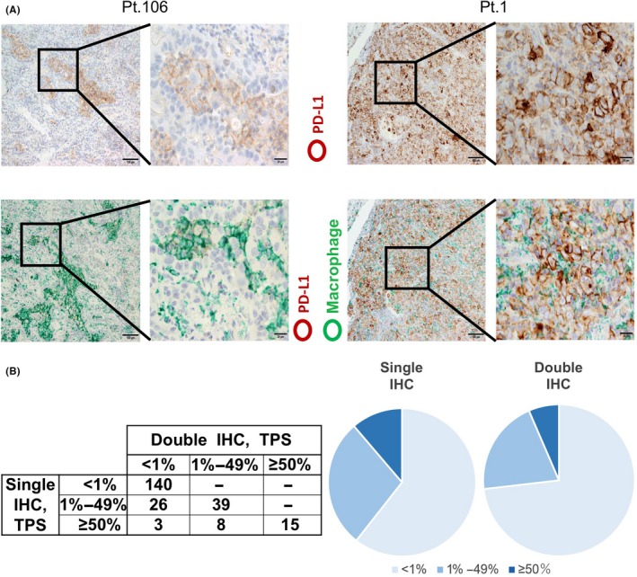
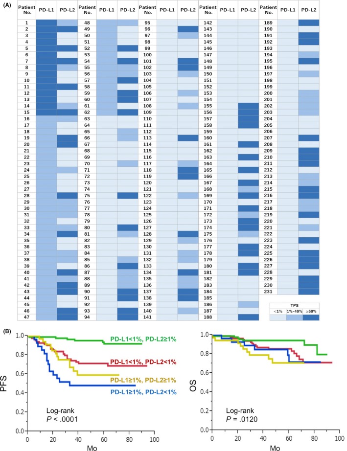
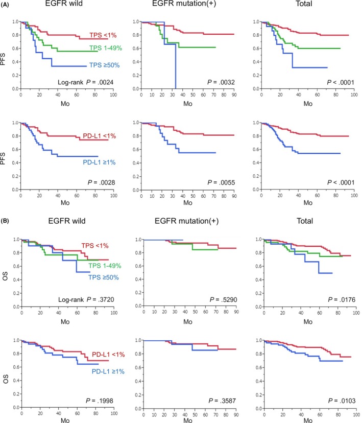
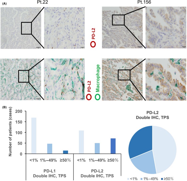
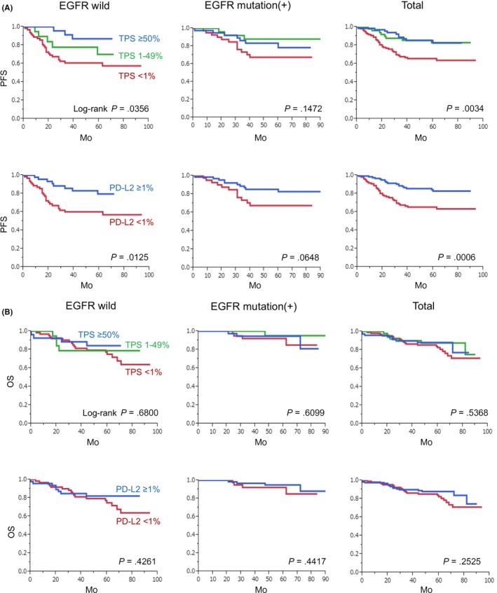
The percentage of programmed death ligand 1 (PD-L1) positivity in cancer cells, named as the tumor proportion score, is considered to be a predictive biomarker for anti-PD-1/PD-L1 therapy in lung cancer. PD-L1 is expressed on not only cancer cells but also on immune cells, including macrophages. Although previous studies related to PD-L1/2 expression in cancer tissues have been generally based on single immunohistochemistry (IHC), in the present study, we attempted to evaluate accurate PD-L1/2 expression in cancer cells in lung adenocarcinoma cells using double IHC to also evaluate macrophages. Of the 231 patients, PD-L1 expression was negative in 169 patients (73.2%), 1%-49% positive in 47 patients (20.3%), and ≥50% positive in 15 patients (6.5%). Interestingly, PD-L1 positivity was decreased when using double IHC compared with the estimation by single IHC. High PD-L1 expression was associated with high-grade cancer cells and in higher stage cancer. PD-L2 was negative in 109 patients (47.2%), 1%-49% positive in 50 patients (21.6%), and ≥50% positive in 72 patients (31.2%). The number of PD-L2-positive patients was increased in cases that had an epidermal growth factor receptor (EGFR) mutation and in lower stage cancer. Thirty-five patients (15.2%) were positive for both PD-L1 and PD-L2, whereas 81 patients (35.1%) were negative for both PD-L1 and PD-L2. Log-rank analysis showed that progression-free survival and overall survival were significantly the longest in the PD-L1-negative and PD-L2-positive groups (P < .0001 and P = .0120). We observed lower PD-L1 or PD-L2 expression in lung adenocarcinoma than previously reported. Double IHC for macrophages may help clinicians to evaluate PD-L1 or PD-L2 expression specifically in cancer cells.
肿瘤细胞程序性死亡配体 1(PD-L1)阳性率(称为肿瘤比例评分)被认为是肺癌抗 PD-1/PD-L1 治疗的预测生物标志物。PD-L1 不仅在癌细胞上表达,而且在包括巨噬细胞在内的免疫细胞上表达。虽然以前与癌症组织中 PD-L1/2 表达相关的研究通常基于单一免疫组化(IHC),但在本研究中,我们试图使用双免疫组化来评估肺腺癌细胞中癌细胞中准确的 PD-L1/2 表达,同时也评估巨噬细胞。在 231 名患者中,169 名(73.2%)患者 PD-L1 表达阴性,47 名(20.3%)患者 PD-L1 表达阳性为 1%-49%,15 名(6.5%)患者 PD-L1 表达阳性为≥50%。有趣的是,与单免疫组化估计相比,使用双免疫组化时 PD-L1 阳性率降低。高 PD-L1 表达与高级别癌细胞和更高分期的癌症有关。109 名(47.2%)患者 PD-L2 阴性,50 名(21.6%)患者 PD-L2 阳性为 1%-49%,72 名(31.2%)患者 PD-L2 阳性为≥50%。在具有表皮生长因子受体(EGFR)突变和更低分期癌症的情况下,PD-L2 阳性患者的数量增加。35 名(15.2%)患者同时 PD-L1 和 PD-L2 阳性,81 名(35.1%)患者同时 PD-L1 和 PD-L2 阴性。对数秩分析显示,PD-L1 阴性和 PD-L2 阳性组的无进展生存期和总生存期明显最长(P<.0001 和 P=.0120)。我们观察到肺腺癌中的 PD-L1 或 PD-L2 表达低于以前的报道。巨噬细胞的双免疫组化可能有助于临床医生专门评估癌细胞中的 PD-L1 或 PD-L2 表达。