Department of Geriatric, Xijing Hospital, the Fourth Military Medical University, 710032, Xi'an, China.
Department of Cardiovascular Surgery, Xijing Hospital, the Fourth Military Medical University, 710032, Xi'an, China.
Cell Death Dis. 2019 Jul 11;10(7):530. doi: 10.1038/s41419-019-1760-5.
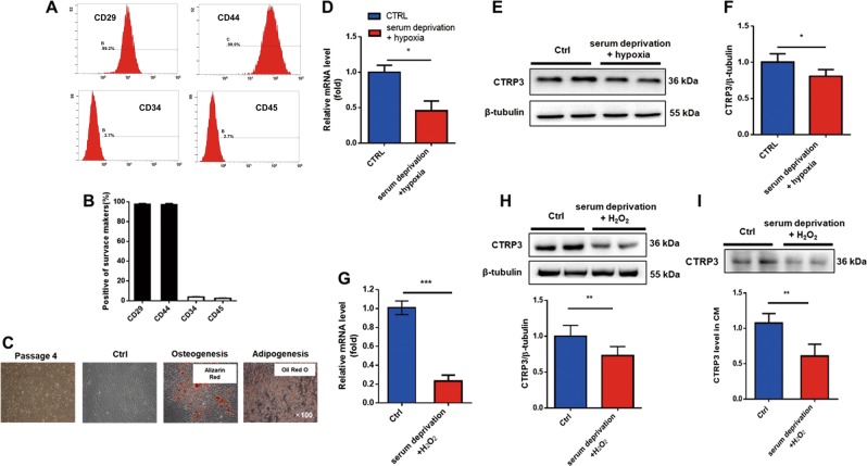
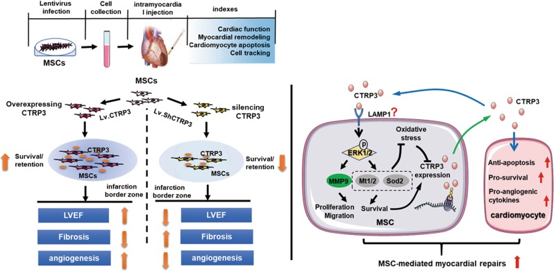
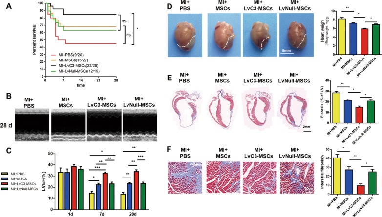
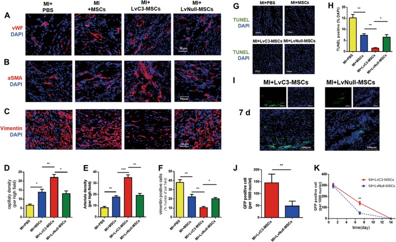
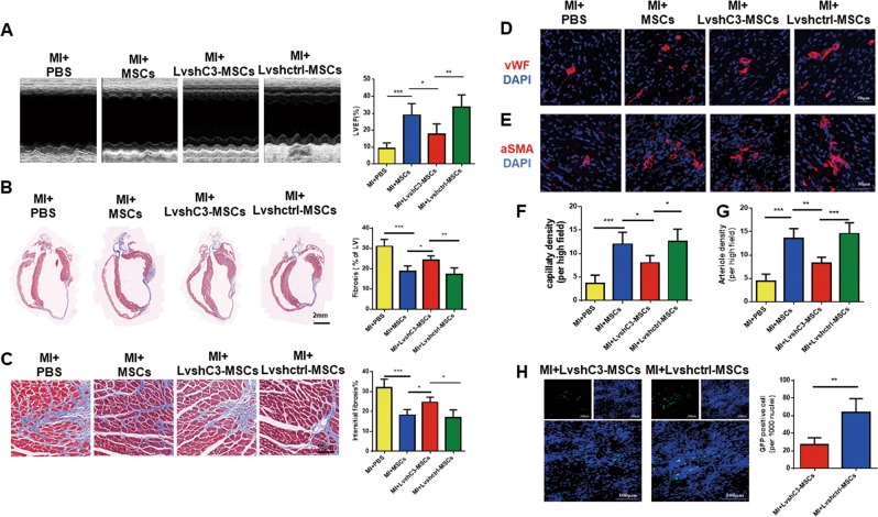
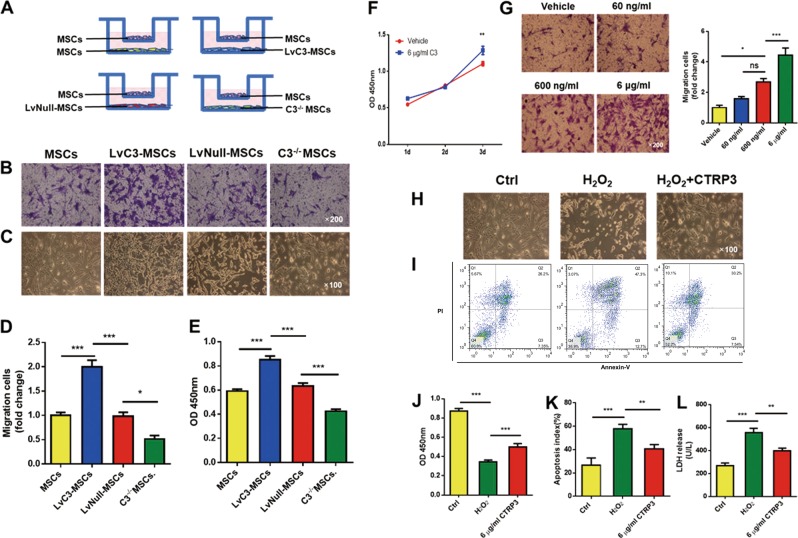
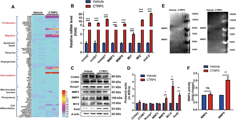
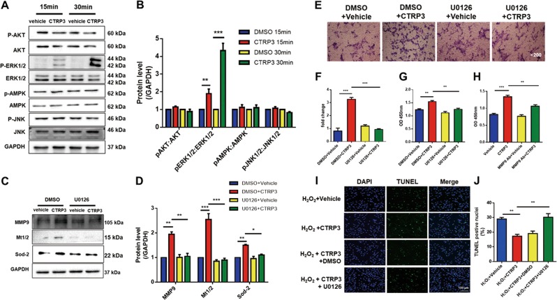
Mesenchymal stromal cells (MSCs) transplantation offers an attractive alternative in myocardial infarctive therapy. However, poor cell engraftment and survival limit their restorative capacity. C1q/tumor necrosis factor-related protein-3 (CTRP3) inhibits reverse remodeling after myocardial infarction (MI) and was found to be secreted by MSCs in our preliminary experiments. We examined whether the overexpression of CTRP3 improved the survival of transplanted MSCs and augmented their efficacy on MI and whether silencing CTRP3 attenuated these effects. For gain-of-function analysis, MSCs overexpressing CTRP3 (LvC3-MSCs), control virus-transfected MSCs (LvNull-MSCs), MSCs alone, or phosphate-buffered saline (PBS) were injected into the peripheral areas of the infarction immediately after coronary artery ligation. For loss-of-function analysis, mice subjected to MI were randomized into groups and administered CTRP3-knockdown MSCs (LvshC3-MSCs), Lvshctrl-MSCs, MSCs, or PBS. Survival rates, cardiac function, and myocardial remodeling in mice were evaluated after 4 weeks. Injection of MSCs or LvNull-MSCs improved the left ventricular ejection fraction, inhibited cardiac fibrosis, and regulated cellular profiles of the infarction border zone 4 weeks after MI compared with those in the PBS group. Furthermore, overexpression of hCTRP3 promoted the efficacy of MSCs in the treatment of MI. However, knocking down CTRP3 impaired that. Coculture experiments confirmed that hCTRP3-enriched conditioned medium (CM) promoted MSCs migration and protected against HO-induced cell damage. Conversely, CM from C3 MSCs (CTRP3 knock out) significantly reduced the migration and antioxidative effects of MSCs. CTRP3 protein alone promoted MSCs proliferation and migration by upregulating matrix metalloproteinase 9 (MMP9) and protecting against oxidation by increasing superoxide dismutase 2 (SOD2) and metallothionein 1/2 (MT1/2) expression; and these effects were blocked by pretreatment with the extracellular signal-regulated kinase (ERK1/2) inhibitor U0126. Overexpression of CTRP3 significantly improved the MSCs-based efficacy on MI by increasing cell survival and retention via a mechanism involving ERK1/2-MMP9 and ERK1/2-SOD2/MT1/2 signaling.
间质基质细胞(MSCs)移植为心肌梗死治疗提供了一种有吸引力的选择。然而,细胞植入和存活不良限制了它们的修复能力。C1q/肿瘤坏死因子相关蛋白-3(CTRP3)抑制心肌梗死后的逆转重构,并且在我们的初步实验中发现其由 MSCs 分泌。我们研究了过表达 CTRP3 是否改善了移植的 MSCs 的存活并增强了它们对心肌梗死的疗效,以及沉默 CTRP3 是否减弱了这些作用。为了进行功能获得分析,将过表达 CTRP3 的 MSCs(LvC3-MSCs)、对照病毒转染的 MSCs(LvNull-MSCs)、单独的 MSCs 或磷酸盐缓冲盐水(PBS)在冠状动脉结扎后立即注射到梗死的外周区域。为了进行功能丧失分析,将发生心肌梗死的小鼠随机分为几组,并给予 CTRP3 敲低 MSCs(LvshC3-MSCs)、Lvshctrl-MSCs、MSCs 或 PBS。在 4 周后评估小鼠的存活率、心功能和心肌重塑。与 PBS 组相比,MSCs 或 LvNull-MSCs 的注射改善了左心室射血分数,抑制了心肌纤维化,并调节了梗死交界区的细胞谱。此外,过表达 hCTRP3 促进了 MSCs 在治疗心肌梗死中的疗效。然而,敲低 CTRP3 则削弱了这种作用。共培养实验证实,富含 hCTRP3 的条件培养基(CM)促进了 MSCs 的迁移并防止了 HO 诱导的细胞损伤。相反,来自 C3 MSCs(CTRP3 敲除)的 CM 显著降低了 MSCs 的迁移和抗氧化作用。CTRP3 蛋白本身通过上调基质金属蛋白酶 9(MMP9)并通过增加超氧化物歧化酶 2(SOD2)和金属硫蛋白 1/2(MT1/2)的表达来促进 MSCs 的增殖和迁移;并且这些作用被细胞外信号调节激酶(ERK1/2)抑制剂 U0126 预处理所阻断。过表达 CTRP3 通过涉及 ERK1/2-MMP9 和 ERK1/2-SOD2/MT1/2 信号通路的机制,通过增加细胞存活和保留来显著改善基于 MSCs 的心肌梗死疗效。