• 文献检索
  • 文档翻译
  • 深度研究
  • 学术资讯
  • Suppr Zotero 插件Zotero 插件
  • 邀请有礼
  • 套餐&价格
  • 历史记录
应用&插件
Suppr Zotero 插件Zotero 插件浏览器插件Mac 客户端Windows 客户端微信小程序
定价
高级版会员购买积分包购买API积分包
服务
文献检索文档翻译深度研究API 文档MCP 服务
关于我们
关于 Suppr公司介绍联系我们用户协议隐私条款
关注我们

Suppr 超能文献

核心技术专利:CN118964589B侵权必究
粤ICP备2023148730 号-1Suppr @ 2026

文献检索

告别复杂PubMed语法,用中文像聊天一样搜索,搜遍4000万医学文献。AI智能推荐,让科研检索更轻松。

立即免费搜索

文件翻译

保留排版,准确专业,支持PDF/Word/PPT等文件格式,支持 12+语言互译。

免费翻译文档

深度研究

AI帮你快速写综述,25分钟生成高质量综述,智能提取关键信息,辅助科研写作。

立即免费体验

在合成神经导管内成像轴突再生。

Imaging axon regeneration within synthetic nerve conduits.

机构信息

Innsbruck Medical University, Department of Anatomy, Histology and Embryology, Division of Neuroanatomy, 6020, Innsbruck, Austria.

VIB-KU Leuven Center for Brain & Disease Research O&N 4, Campus Gasthuisberg, 3000, Leuven, Belgium.

出版信息

Sci Rep. 2019 Jul 12;9(1):10095. doi: 10.1038/s41598-019-46579-w.

DOI:10.1038/s41598-019-46579-w
PMID:31300753
原文链接:https://pmc.ncbi.nlm.nih.gov/articles/PMC6626049/
Abstract

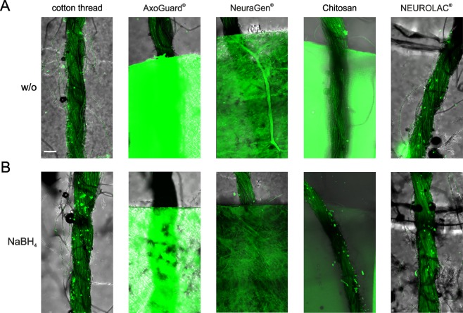
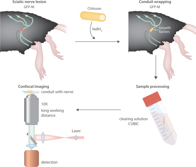
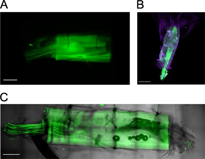
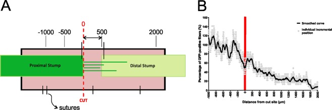
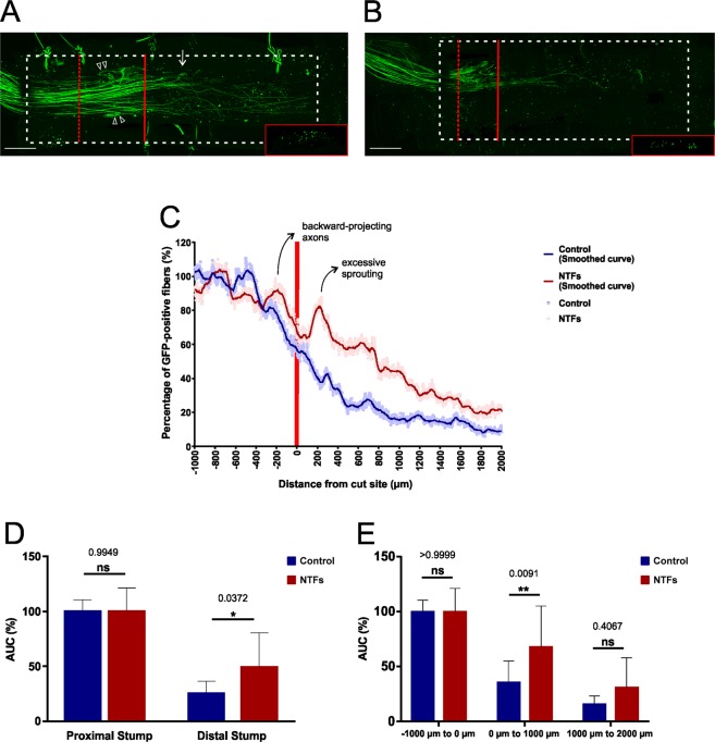
While axons within the central nervous system (CNS) do not regenerate following injury, those in the peripheral nervous system (PNS) do, although not in a clinically satisfactory manner as only a small proportion of axons exhibit long-distance regeneration. Moreover, functional recovery is hampered by excessive axonal sprouting and aberrant reinnervation of target tissue. In order to investigate the mechanisms governing the regrowth of axons following injury, previous studies have used lesion paradigms of peripheral nerves in rat or mouse models, and reagents or cells have been administered to the lesion site through nerve conduits, aiming to improve early-stage regeneration. Morphological analysis of such in vivo experiments has however been limited by the incompatibility of synthetic nerve conduits with existing tissue-clearing and imaging techniques. We present herein a novel experimental approach that allows high-resolution imaging of individual axons within nerve conduits, together with quantitative assessment of fiber growth. We used a GFP-expressing mouse strain in a lesion model of the sciatic nerve to describe a strategy that combines nerve clearing, chemical treatment of chitosan nerve conduits, and long working distance confocal microscopy with image processing and analysis. This novel experimental setup provides a means of documenting axon growth within the actual conduit during the critical initial stage of regeneration. This will greatly facilitate the development and evaluation of treatment regimens to improve axonal regeneration following nerve damage.

摘要

虽然中枢神经系统(CNS)中的轴突在受伤后不会再生,但周围神经系统(PNS)中的轴突会再生,尽管其方式在临床上并不令人满意,因为只有一小部分轴突表现出长距离再生。此外,轴突过度发芽和靶组织的异常再支配会阻碍功能恢复。为了研究损伤后轴突再生的调控机制,以前的研究使用了大鼠或小鼠模型中的周围神经损伤范例,并通过神经导管向损伤部位给予试剂或细胞,旨在改善早期再生。然而,这种体内实验的形态学分析受到合成神经导管与现有组织清除和成像技术不兼容的限制。我们在此提出了一种新的实验方法,允许在神经导管内对单个轴突进行高分辨率成像,并对纤维生长进行定量评估。我们使用了一种 GFP 表达的小鼠品系在坐骨神经损伤模型中,描述了一种结合神经清除、壳聚糖神经导管化学处理以及长工作距离共聚焦显微镜与图像处理和分析的策略。这种新的实验装置提供了一种在再生的关键初始阶段在实际导管内记录轴突生长的方法。这将极大地促进改善神经损伤后轴突再生的治疗方案的开发和评估。

https://cdn.ncbi.nlm.nih.gov/pmc/blobs/b814/6626049/d893af93ec4a/41598_2019_46579_Fig5_HTML.jpg
https://cdn.ncbi.nlm.nih.gov/pmc/blobs/b814/6626049/cc84f625936b/41598_2019_46579_Fig1_HTML.jpg
https://cdn.ncbi.nlm.nih.gov/pmc/blobs/b814/6626049/547651033cc9/41598_2019_46579_Fig2_HTML.jpg
https://cdn.ncbi.nlm.nih.gov/pmc/blobs/b814/6626049/6d3fa87e0bbe/41598_2019_46579_Fig3_HTML.jpg
https://cdn.ncbi.nlm.nih.gov/pmc/blobs/b814/6626049/1814c8754b2e/41598_2019_46579_Fig4_HTML.jpg
https://cdn.ncbi.nlm.nih.gov/pmc/blobs/b814/6626049/d893af93ec4a/41598_2019_46579_Fig5_HTML.jpg

相似文献

1
Imaging axon regeneration within synthetic nerve conduits.在合成神经导管内成像轴突再生。
Sci Rep. 2019 Jul 12;9(1):10095. doi: 10.1038/s41598-019-46579-w.
2
Permeable guidance channels containing microfilament scaffolds enhance axon growth and maturation.包含微丝支架的可渗透引导通道可促进轴突生长和成熟。
J Biomed Mater Res A. 2005 Nov 1;75(2):374-86. doi: 10.1002/jbm.a.30432.
3
Applications of Proteomics to Nerve Regeneration Research蛋白质组学在神经再生研究中的应用
4
A comparison of the performance of mono- and bi-component electrospun conduits in a rat sciatic model.单组分和双组分电纺导管在大鼠坐骨模型中的性能比较。
Biomaterials. 2014 Oct;35(32):8970-82. doi: 10.1016/j.biomaterials.2014.07.010. Epub 2014 Jul 29.
5
Electrospun silk-polyaniline conduits for functional nerve regeneration in rat sciatic nerve injury model.静电纺丝丝-聚苯胺导管在大鼠坐骨神经损伤模型中的功能神经再生作用。
Biomed Mater. 2017 Aug 17;12(4):045025. doi: 10.1088/1748-605X/aa7802.
6
Use of hybrid chitosan membranes and N1E-115 cells for promoting nerve regeneration in an axonotmesis rat model.在轴突断裂大鼠模型中使用混合壳聚糖膜和N1E-115细胞促进神经再生
Biomaterials. 2008 Nov;29(33):4409-19. doi: 10.1016/j.biomaterials.2008.07.043. Epub 2008 Aug 23.
7
Comprehensive evaluation of peripheral nerve regeneration in the acute healing phase using tissue clearing and optical microscopy in a rodent model.在啮齿动物模型中,使用组织透明化和光学显微镜对急性愈合期周围神经再生进行综合评估。
PLoS One. 2014 Apr 8;9(4):e94054. doi: 10.1371/journal.pone.0094054. eCollection 2014.
8
PDLLA/chondroitin sulfate/chitosan/NGF conduits for peripheral nerve regeneration.聚(D,L-乳酸)/硫酸软骨素/壳聚糖/神经生长因子导管修复周围神经损伤。
Biomaterials. 2011 Jul;32(20):4506-16. doi: 10.1016/j.biomaterials.2011.02.023. Epub 2011 Mar 11.
9
Microwave irradiated collagen tubes as a better matrix for peripheral nerve regeneration.微波辐照胶原管作为周围神经再生的更佳基质。
Brain Res. 2005 Jun 7;1046(1-2):55-67. doi: 10.1016/j.brainres.2005.03.022.
10
The pros and cons of growth factors and cytokines in peripheral axon regeneration.生长因子和细胞因子在外周轴突再生中的利弊。
Int Rev Neurobiol. 2013;108:137-71. doi: 10.1016/B978-0-12-410499-0.00006-X.

引用本文的文献

1
Advanced development of conductive biomaterials for enhanced peripheral nerve regeneration: a review.用于增强周围神经再生的导电生物材料的研究进展:综述
RSC Adv. 2025 Apr 22;15(17):12997-13009. doi: 10.1039/d5ra01107h.
2
Role of transforming growth factor-β in peripheral nerve regeneration.转化生长因子-β在外周神经再生中的作用。
Neural Regen Res. 2024 Feb;19(2):380-386. doi: 10.4103/1673-5374.377588.
3
Tissue optical clearing for neural regeneration research.用于神经再生研究的组织光学透明化

本文引用的文献

1
Enhancing the Outcome of Traumatic Sensory Nerve Lesions of the Hand by Additional Use of a Chitosan Nerve Tube in Primary Nerve Repair: A Randomized Controlled Bicentric Trial.在初次神经修复时附加使用壳聚糖神经导管增强手部创伤性感觉神经损伤的疗效:一项随机对照双中心试验。
Plast Reconstr Surg. 2018 Aug;142(2):415-424. doi: 10.1097/PRS.0000000000004574.
2
RTF: a rapid and versatile tissue optical clearing method.RTF:一种快速且多功能的组织光学透明化方法。
Sci Rep. 2018 Jan 31;8(1):1964. doi: 10.1038/s41598-018-20306-3.
3
Whole-Brain Microscopy Meets In Vivo Neuroimaging: Techniques, Benefits, and Limitations.
Neural Regen Res. 2023 Sep;18(9):1940-1941. doi: 10.4103/1673-5374.363827.
4
Biodegradable Nerve Guide with Glial Cell Line-Derived Neurotrophic Factor Improves Recovery After Facial Nerve Injury in Rats.可生物降解神经引导物联合胶质细胞源性神经营养因子促进大鼠面神经损伤后的恢复。
Facial Plast Surg Aesthet Med. 2023 Nov-Dec;25(6):478-486. doi: 10.1089/fpsam.2022.0346. Epub 2023 Mar 6.
5
Glial Cell Line-Derived Neurotrophic Factor-Loaded CMCht/PAMAM Dendrimer Nanoparticles for Peripheral Nerve Repair.用于周围神经修复的载有胶质细胞源性神经营养因子的羧甲基壳聚糖/聚酰胺-胺树枝状大分子纳米颗粒
Pharmaceutics. 2022 Nov 8;14(11):2408. doi: 10.3390/pharmaceutics14112408.
6
Bioactive Nanofiber-Based Conduits in a Peripheral Nerve Gap Management-An Animal Model Study.基于生物活性纳米纤维的周围神经间隙管理导管:一项动物模型研究。
Int J Mol Sci. 2021 May 25;22(11):5588. doi: 10.3390/ijms22115588.
7
Ten-eleven translocation methylcytosine dioxygenase 3-loaded microspheres penetrate neurons in vitro causing active demethylation and neurite outgrowth.Ten-eleven 易位甲基胞嘧啶双加氧酶 3 负载微球在体外穿透神经元,导致活性去甲基化和神经突生长。
J Tissue Eng Regen Med. 2021 May;15(5):463-474. doi: 10.1002/term.3185. Epub 2021 Mar 18.
8
Collaborating by courier, imaging by mail.快递协作,邮件成像。
EMBO Rep. 2020 Jan 7;21(1):e49755. doi: 10.15252/embr.201949755. Epub 2019 Dec 16.
全脑显微镜与活体神经成像:技术、优势及局限性
Mol Imaging Biol. 2017 Feb;19(1):1-9. doi: 10.1007/s11307-016-0988-z.
4
Autograft Substitutes: Conduits and Processed Nerve Allografts.自体移植替代物:导管与处理过的同种异体神经移植物。
Hand Clin. 2016 May;32(2):127-40. doi: 10.1016/j.hcl.2015.12.012.
5
ACT-PRESTO: Rapid and consistent tissue clearing and labeling method for 3-dimensional (3D) imaging.ACT-PRESTO:用于三维(3D)成像的快速且一致的组织透明化和标记方法。
Sci Rep. 2016 Jan 11;6:18631. doi: 10.1038/srep18631.
6
Schwann Cell Expressed Nogo-B Modulates Axonal Branching of Adult Sensory Neurons Through the Nogo-B Receptor NgBR.施万细胞表达的Nogo-B通过Nogo-B受体NgBR调节成年感觉神经元的轴突分支。
Front Cell Neurosci. 2015 Nov 23;9:454. doi: 10.3389/fncel.2015.00454. eCollection 2015.
7
Chitosan-film enhanced chitosan nerve guides for long-distance regeneration of peripheral nerves.壳聚糖膜增强的壳聚糖神经导管促进周围神经的长距离再生。
Biomaterials. 2016 Jan;76:33-51. doi: 10.1016/j.biomaterials.2015.10.040. Epub 2015 Oct 21.
8
Loss of GPR3 reduces the amyloid plaque burden and improves memory in Alzheimer's disease mouse models.GPR3 的缺失减少了阿尔茨海默病小鼠模型中的淀粉样斑块负担并改善了记忆。
Sci Transl Med. 2015 Oct 14;7(309):309ra164. doi: 10.1126/scitranslmed.aab3492.
9
Trends in the design of nerve guidance channels in peripheral nerve tissue engineering.周围神经组织工程中神经引导通道设计的发展趋势。
Prog Neurobiol. 2015 Aug;131:87-104. doi: 10.1016/j.pneurobio.2015.06.001. Epub 2015 Jun 18.
10
GDNF preconditioning can overcome Schwann cell phenotypic memory.胶质细胞源性神经营养因子预处理可克服雪旺细胞表型记忆。
Exp Neurol. 2015 Mar;265:1-7. doi: 10.1016/j.expneurol.2014.12.003. Epub 2014 Dec 11.