Department of Neurology and Neurosurgery, Dick White Referrals, Six Mile Bottom, UK.
Department of Diagnostic Imaging, Dick White Referrals, Six Mile Bottom, UK.
Vet Rec. 2019 Sep 14;185(10):306. doi: 10.1136/vr.105243. Epub 2019 Jul 15.
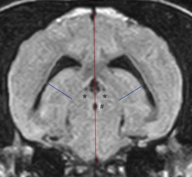
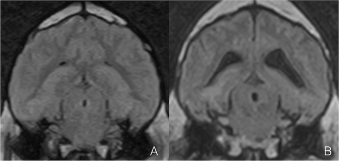
Age-related hippocampal formation (HF) atrophy has been documented on MRI studies using volumetric analysis and visual rating scales.This retrospective cross-sectional study aimed to compare linear MRI measurements of the HF between young (1-3 years) and old (>10 years) non-brachycephalic dogs, with normal brain anatomy and cerebrospinal fluid (CSF) analysis. Right and left hippocampal formation height (HFH), height of the brain (HB) and mean HFH/HB ratio were measured by two observers on a transverse T2 fluid-attenuated inversion recovery sequence containing rostral colliculi and mesencephalic aqueduct.119 MRI studies were enrolled: 75 young and 44 old dogs. Left and right HFH were greater (p<0.0001) in young, while HB was greater in old dogs (p=0.024). Mean HFH/HB ratio was 15.66 per cent and 18.30 per cent in old and young dogs (p<0.0001). No differences were found comparing measurements between epileptic and non-epileptic dogs.Old dogs have a greater HB; this may represent the different study populations or a statistical phenomenon. Ageing affects HF linear measurements. A reduction of mean HFH/HB ratio between 18.30 per cent and 15.66 per cent should be considered a physiological age-related process of the canine lifespan. The use of mean HFH/HB ratio could be considered for quantifying brain atrophy in elderly dogs.
MRI 研究通过容积分析和视觉评分量表已经证实了与年龄相关的海马结构(HF)萎缩。这项回顾性的横断面研究旨在比较正常脑解剖结构和脑脊液(CSF)分析的年轻(1-3 岁)和年长(>10 岁)非短头犬的 HF 的线性 MRI 测量值。两名观察者在包含颅神经和中脑导水管的横向 T2 液体衰减反转恢复序列上测量了右侧和左侧 HF 高度(HFH)、脑高(HB)和平均 HFH/HB 比值。共纳入 119 项 MRI 研究:75 只幼犬和 44 只老年犬。幼犬的左、右 HFH 更大(p<0.0001),而老年犬的 HB 更大(p=0.024)。老年犬和幼犬的平均 HFH/HB 比值分别为 15.66%和 18.30%(p<0.0001)。在比较癫痫和非癫痫犬的测量值时,未发现差异。老年犬的 HB 较大;这可能代表不同的研究人群或统计现象。年龄的增长会影响 HF 的线性测量值。平均 HFH/HB 比值从 18.30%降低至 15.66%应被视为犬寿命的生理性年龄相关过程。在老年犬中,使用平均 HFH/HB 比值可以考虑用于量化脑萎缩。