Department of Nanoengineering, University of California San Diego, La Jolla, CA, USA.
School of Medicine, University of California San Diego, La Jolla, CA, USA.
Sci Rep. 2019 Jul 16;9(1):10279. doi: 10.1038/s41598-019-46311-8.
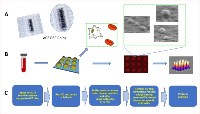
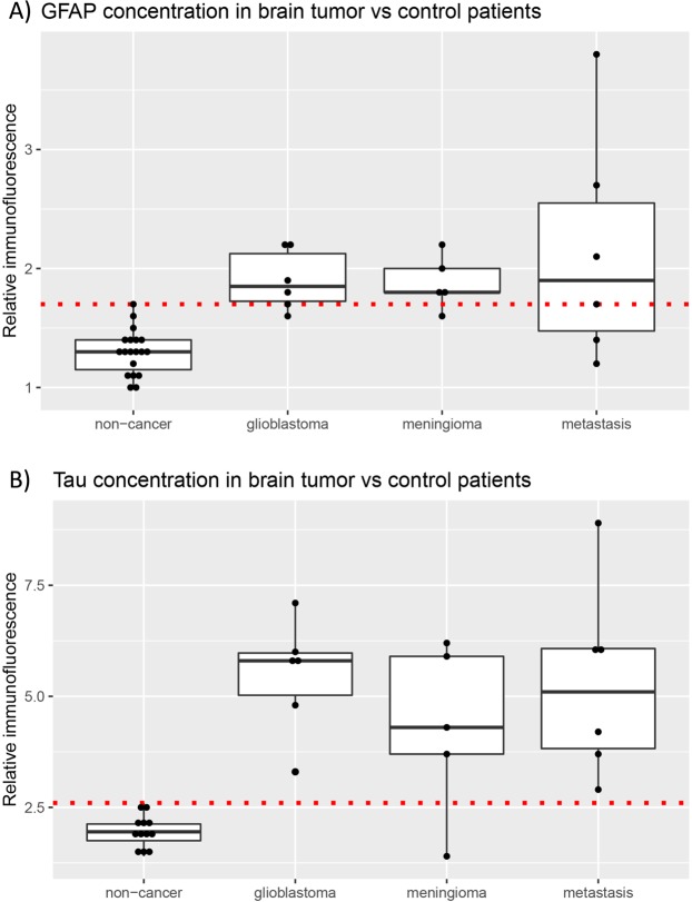
Extracellular vesicles (EVs) are small, membrane-bound particles released by all cells that have emerged as an attractive biomarker platform. We study the utility of a dielectrophoretic (DEP) micro-chip device for isolation and characterization of EVs derived from plasma specimens from patients with brain tumors. EVs were isolated by DEP chip and subjected to on-chip immunofluorescence (IF) staining to determine the concentration of glial fibrillary acidic protein (GFAP) and Tau. EVs were analyzed from the plasma samples isolated from independent patient cohorts. Glioblastoma cell lines secrete EVs enriched for GFAP and Tau. These EVs can be efficiently isolated using the DEP platform. Application of DEP to clinical plasma samples afforded discrimination of plasma derived from brain tumor patients relative to those derived from patients without history of brain cancer. Sixty-five percent (11/17) of brain tumor patients showed higher EV-GFAP than the maximum observed in controls. Ninety-four percent (16/17) of tumor patients showed higher EV-Tau than the maximum observed in controls. These discrimination thresholds were applied to plasma isolated from a second, independent cohort of 15 glioblastoma patients and 8 controls. For EV-GFAP, we observed 93% sensitivity, 38% specificity, 74% PPV, 75% NPV, and AUC of 0.65; for EV-Tau, we found 67% sensitivity, 75% specificity 83% PPV, 55% NPV, and AUC of 0.71 for glioblastoma diagnosis. This proof-of-principle study provides support for DEP-IF of plasma EVs for diagnosis of glioblastoma.
细胞外囊泡 (EVs) 是所有细胞释放的小的、膜结合的颗粒,已成为有吸引力的生物标志物平台。我们研究了介电泳 (DEP) 微芯片设备在分离和鉴定源自脑肿瘤患者血浆标本的 EVs 方面的应用。通过 DEP 芯片分离 EVs,并进行芯片上免疫荧光 (IF) 染色,以确定神经胶质纤维酸性蛋白 (GFAP) 和 Tau 的浓度。从独立患者队列分离的血浆样本中分析 EVs。神经胶质瘤细胞系分泌富含 GFAP 和 Tau 的 EVs。这些 EVs可以使用 DEP 平台高效分离。DEP 在临床血浆样本中的应用能够区分源自脑肿瘤患者的血浆与源自无脑瘤病史患者的血浆。65%(11/17)的脑肿瘤患者的 EV-GFAP 高于对照组中观察到的最大值。94%(16/17)的肿瘤患者的 EV-Tau 高于对照组中观察到的最大值。这些区分阈值应用于来自第二组 15 名胶质母细胞瘤患者和 8 名对照组的独立血浆样本。对于 EV-GFAP,我们观察到 93%的敏感性、38%的特异性、74%的阳性预测值、75%的阴性预测值和 0.65 的 AUC;对于 EV-Tau,我们发现 67%的敏感性、75%的特异性、83%的阳性预测值、55%的阴性预测值和 0.71 的 AUC 用于胶质母细胞瘤诊断。这项初步研究为 DEP-IF 分析血浆 EVs 用于胶质母细胞瘤诊断提供了支持。